


Fig. 2.
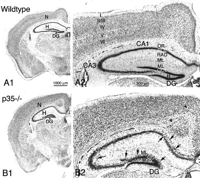
A1, B1, Coronal brain sections from wild-type (A1) and p35−/− knock-out (B1) mice, stained with cresyl violet. Neocortex (N), hippocampus (H), and dentate gyrus (DG) are indicated. A2, Higher magnification of neocortex and hippocampus from a wild-type mouse shows normal neocortical laminae (I–VI) and hippocampal subregionsCA1 and CA3 with s. oriens (OR) and radiatum (RAD), pyramidal cell layer (PCL), molecular layer (ML), and dentate gyrus (DG) with hilus (H), granule cell layer (GCL), and molecular layer (ML).B2, Coronal section of neocortex and hippocampus of a p35−/− mouse demonstrates heterotopic pyramidal neurons in CA1/CA3 (arrows) and dispersed dentate granule cells localized within the inner molecular layer (ML;arrows). Note the abnormal cortical lamination, the presence of aberrant fiber fascicles within the neocortex (arrowheads), and the absence of callosal fibers crossing the midline (white arrow). Scale bars:A1, B1, 1000 μm; A2,B2, 300 μm.