


Fig. 2.
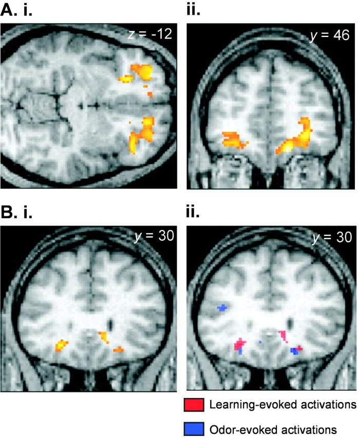
Valence-independent learning. A, Rostral orbitofrontal cortex. The SPM (threshold, p< 0.001) is overlaid on the axial (i) and coronal (ii) sections of a subject's normalized T1-weighted scan and depicts extensive bilateral activations in rostral areas of orbitofrontal cortex. In this and all subsequent figures, the right side of the brain corresponds to the right side of the image. B, Caudal orbitofrontal cortex.i, Neural activations in caudal orbitofrontal cortex are superimposed on a normalized T1-weighted coronal image (threshold,p < 0.001). ii, The learning-evoked activations depicted in i are illustrated inred (threshold, p < 0.001) and overlap the odor-evoked activations derived from the companion study (Gottfried et al., 2002), which are shown in blue(threshold, p < 0.005).