


Fig. 3.
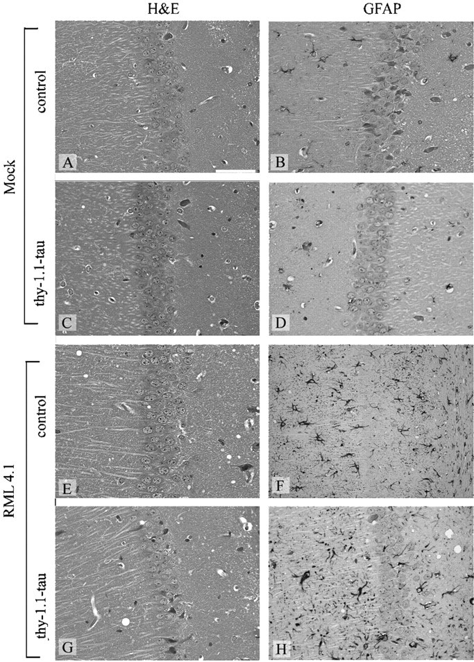
Histopathological findings in hippocampi of mice. Hematoxylin and eosin-stained (A, C, E, G) and GFAP-stained (B, D, F, H) brain sections of thy-1.1 tau (C, D, G, H) and control mice (A, B, E, F) after intranerval inoculation of scrapie prions (E–H) and mock inoculum (A–D). Spongiform changes and intense, diffuse gliosis were visible in brains of infected thy-1.1-tau and control mice. Scale bar, 50 μm.