


Fig. 3.
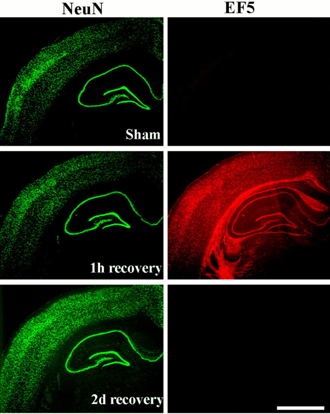
Immunohistochemistry for the in vivo hypoxia marker EF5 showing weak signal (red) in the sham-operated rat brain but strong signal at 1 hr of recovery from cardiac arrest and resuscitation. At 48 hr of recovery, EF5 adduct binding was comparable with the sham-control. The same sections were immunostained for the neuronal marker NeuN (green). The nitroimidazole compound EF5 was injected intravenously as described in Materials and Methods. Scale bar, 1 mm.