


Figure 1.
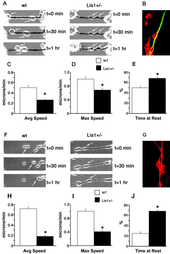
Lis1+/- cerebellar granule neurons display motility defects in both glia- and neurite-guided migration assays. A, Wild-type neurons cover a longer distance when migrating on glia than Lis1+/- neurons, as shown by displacement of the cell body (arrow) over the period of 1 hr (video-enhanced phase-contrast microscopy, 30 min between planes). B, Glia-guided assay cultures stained with βIII-tubulin (neurons; red) and GFAP (glia; green) antibodies. C, Average speed of Lis1+/- neurons (n = 17) compared with wt neurons (n = 43). D, The maximal speed of neurons was decreased in Lis1+/- neurons, whereas the percentage of time at rest (E) was increased. F, In reaggregate cultures, neurons cover a longer distance when migrating on neurites than Lis1+/-, neurons as shown by displacement of the cell body (arrow) over the period of 1 hr (video-enhanced phase-contrast microscopy, 30 min between planes). G, Dual labeling with βIII-tubulin (neurons; red) and GFAP (glia; green) antibodies show reaggregate cultures are >95% neurons. Average speed (H), maximal velocity (I), and time spent at rest (J) are all negatively affected in Lis1+/- neurons on neurites (n = 12) compared with wt neurons (n = 21). *p < 0.01.