


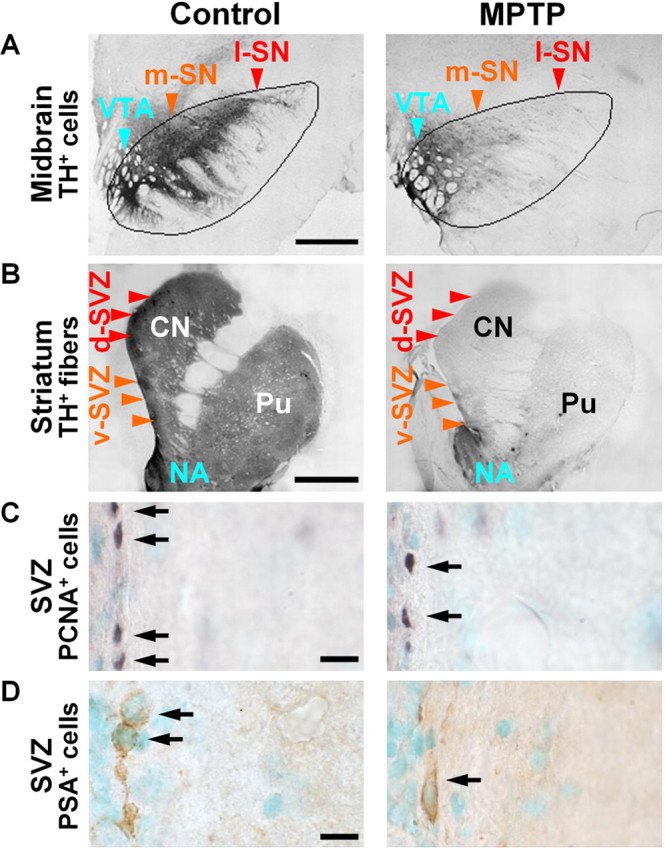
Dopaminergic denervation and reduction in the number of precursor cells in the SVZ of MPTP-treated aged macaques (20–25 years). A, B, Dopaminergic cell bodies and fibers were visualized by immunolabeling TH in the ventral midbrain (A) and striatum (B) in control and MPTP-treated macaques. VTA, Ventral tegmental area; SN, pars compacta of the substantia nigra; m, medial; l, lateral; d, dorsal; v, ventral; CN, caudate nucleus; Pu, putamen; NA, nucleus accumbens. Note that degeneration of the l-SN (red) projecting to the d-SVZ (red) is severe in MPTP-treated monkeys, whereas the m-SN (orange) projecting to the v-SVZ (orange) and the VTA (blue) projecting to the NA (blue) are only moderately or mildly affected, respectively. Scale bars: A, B, 2 mm. C, D, Representative photomicrographs showing PCNA+ proliferating cells (C, arrows) and PSA-NCAM+ migrating neuroblasts (D, arrows) in the SVZ of a control and an MPTP monkey. Nuclei are counterstained with methyl green. Scale bars: C, D, 10 μm.