


Figure 4.
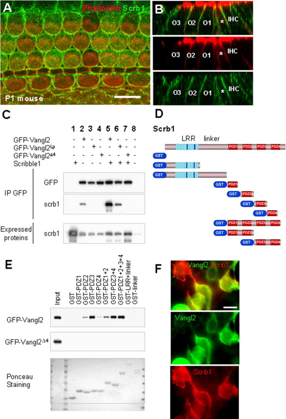
Scrb1 and Vangl2 physically interact, and Scrb1 is required for Vangl2 asymmetry. A, Lumenal surface of a P1 mouse cochlea labeled with an antibody against Scrb1 (green) indicates widespread expression of the protein at the membranes of all the cells in the OC. B, Confocal Z-stack cross-section of a P1 mouse cochlea labeled as in A. Top illustrates merged image, and middle and bottom indicate individual phalloidin (red) and Scrb1 (green) channels. Scrb1 is distributed along the entire basolateral membranes of the IHCs and OHCs (numbered) as well as the IPs (asterisk). C, Immunoprecipitation of Scrb1 with Vangl2. HEK293 cells were transfected with Scrb1 and GFP-Vangl2 or GFP-Vangl2Lp constructs, and proteins were immunoprecipitated with an anti-GFP antibody. The membranes were then probed with our anti-Scrb1 antibody. The presence of a band indicates that Vangl2 and Vangl2Lp can interact with Scrb1 (lanes 5, 6), but removal of the PDZ binding domain at the C terminus of Vangl2 (Vangl2Δ4) prevents this interaction (lane 7). The band in column 2 represents endogenously expressed Scrb1 in HEK293 cells. D, Structure of mammalian Scrb1, with an LRR near the N terminus, a linker region, and four PDZ domains at the C terminus. Beneath the full structure is a graphical list of the GST constructs used in the GST pull-down assay in E. E, GST pull downs indicate that Vangl2 can interact with PDZ2, PDZ3, or PDZ4, as well as other combinations of PDZ domains from Scrb1, with the strongest interaction with PDZ2 and PDZ3. In contrast, Vangl2 does not interact with the LRR or linker domains. Mutation of Vangl2 on serine S464 leads to a strong reduction in Scrb1 binding, and mutation of the last four amino acids of Vangl2 (Vangl2Δ4) completely suppresses this binding. F, Immunolocalization of Vangl2 and Scrb1 in HEK293 cells. HEK293 cells were transfected with Scrb1 (red) and GFP-Vangl2 (green) constructs, and proteins were processed for immunocytochemistry with anti-GFP and anti-Scrb1 antibodies. The two proteins colocalize strongly when coexpressed in the cells. Scale bar, 10 μm.