


Figure 8.
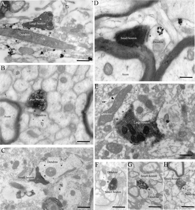
Ultrastructure of large and small boutons in TRN. A, B, EM photomicrographs showing large labeled boutons synapsing on PV+ dendrites (black dots). C, EM photomicrograph showing a small labeled bouton synapsing on a PV+ dendrite. D, Small bouton synapsing on a PV+ dendrite. E, Large bouton synapsing on a GABA+ dendrite (black dots). F–H, Small boutons synapsing on GABA+ dendrites. Asterisks indicate postsynaptic densities. Dendrites labeled for PV or GABA contain gold particles (black dots). Scale bars: A–C, E–H, 1 μm; D, 0.5 μm.