


Figure 8.
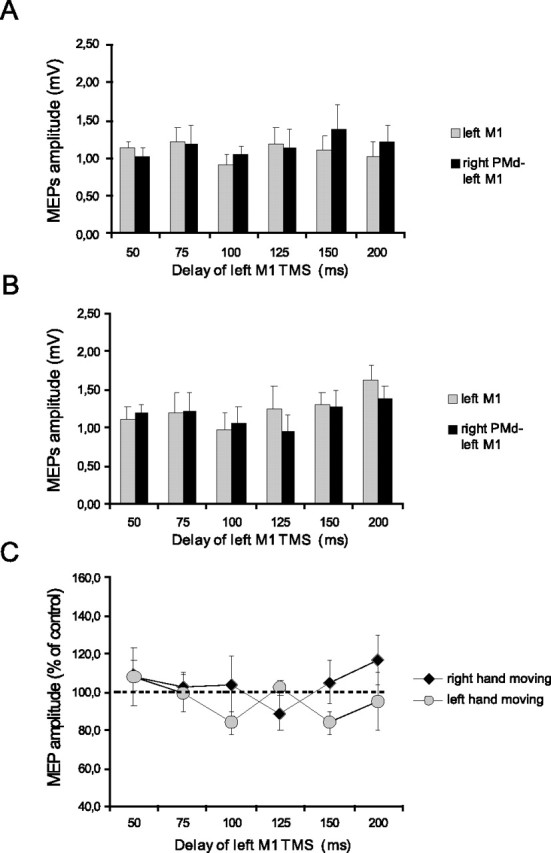
Effects of low-intensity (80% AMT) right PMd conditioning on contralateral M1 excitability at different delays after the cue signal. A, B, MEP amplitudes recorded from the right FDI during either condition (single TMS or ppTMS) at different time points at which the left (A) or the right (B) hand was selected for movement. C, Mean percentage values of the change on MEP amplitudes during right PMd conditioning. No significant effect was observed during the task. Error bars indicate SEM.