


Figure 2.
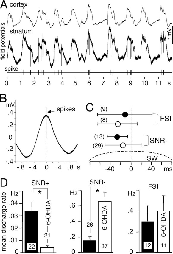
Spontaneous discharge activity of striatal neurons during slow waves. A, Cortical and striatal field potentials were recorded simultaneously with the discharge activity of one SNR− neuron in a 6-OHDA-lesioned rat. Notice that the striatal field potential closely followed the cortical one and that action potentials only occurred during the maximum of the field potentials. B, Temporal relationship between discharge activity and striatal slow waves. The discharge activity (174 spikes) was recorded simultaneously with the field potential during a continuous period of SW+ state (267 s). Sections of striatal field potential recordings centered on every spike were averaged together. On average, this SNR− neuron recorded in a 6-OHDA-lesioned rat discharged after the maximum of the slow waves. C, Temporal relationship between the striatal SW and the discharge activity of FSI and presumed striatopallidal neurons (SNR−) recorded in sham-operated rats (filled circles) and in 6-OHDA lesioned rats (open circles). The number of neurons studied is indicated in parentheses. Error bars indicate mean ± SD. All neurons discharged closely to the maximum of the striatal slow wave. D, Mean discharge activities of striatonigral neurons (SNR+), presumed striatopallidal neurons (SNR−), and FSIs compared in sham-operated rats (filled bars) and in 6-OHDA-lesioned rats (open bars). Bar histograms indicate mean ± SEM (unpaired t test, *p ≤ 0.0019). The number of neurons studied is indicated inside or above bars. The dopaminergic depletion strongly decreased the discharge activity of striatonigral neurons and enhanced that of striatopallidal neurons.