


Figure 8.
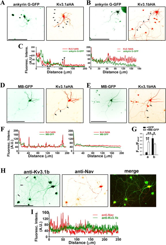
Ankyrin G differentially regulates polarized targeting of Kv3.1 splice variants. A–G, Hippocampal neurons at 7 DIV were cotransfected with ankyrin G and Kv3.1 constructs, fixed, and stained with anti-HA antibodies under permeabilized conditions. A, Kv3.1aHA (red) did not colocalize with ankyrin G-GFP (green) in axons. B, Kv3.1bHA (red) colocalized with ankyrin G-GFP (green) in proximal axons. C, The fluorescence intensity profiles along the axons in A and B indicated by arrows. Arrowheads, The fluorescence peaks attributable to crossing dendrites. D, E, The levels of Kv3.1aHA (D) and Kv3.1bHA (E) in axons markedly increased and modestly decreased, respectively, in the presence of MB–GFP (green). F, The fluorescence intensity profiles along the axons in D and E indicated by arrows. G, Summary of polarized targeting of Kv3.1aHA and Kv3.1bHA in the presence of GFP and MB–GFP. Faxon/Fdendrite is average fluorescence intensity in axons/average fluorescence intensity in dendrites. H, Endogenous Kv3.1b channels were localized to distal axons (arrows), in contrast to the localization of endogenous Nav channels in proximal axons. Hippocampal neurons at 30 DIV were costained with polyclonal anti-Kv3.1b (green) and monoclonal anti-pan-Nav channel (red) antibodies. It is important to note that Kv3.1b also localizes in soma and proximal dendrites. I, Fluorescence intensity profiles along the axon indicated by arrows in H. Distance 0, Soma. Scale bars, 100 μm. Student's t test used in the comparison of two groups. *p < 0.01; **p < 0.0001.