


Figure 10.
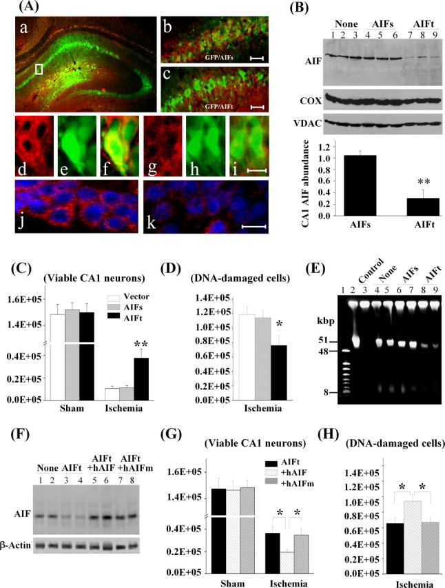
AIF knockdown is neuroprotective in CA1 against transient global ischemia. A, CA1 was coinfected for 14 d with AAV–GFP and the AIF-targeting sequence AAV–AIFt (a, c) or the AIF scramble sequence AAV–AIFs (b). d–f show double-label immunofluorescence of AIF (red) and GFP (green) in AAV–AIFs/AAV–GFP-infected CA1 neurons; g–i show reduced AIF expression (red) in AAV–AIFt/AAV–GFP-infected CA1 neurons. j and k show double-label immunofluorescence of AIF (red) and Hoechst 33258 (blue) in AAV–AIFs- and AAV–AIFt-infected CA1, respectively. B, Western blots show the expression levels of AIF in CA1, either non-infected (lanes 1–3) or at 14 d after infection with AAV–AIFs (lanes 4–6) or AAV–AIFt (lanes 7–9). The graph illustrates the relative changes of AIF expression in CA1 after AAV–AIFt infection. **p < 0.01 versus AAV–AIFs-infected brains; n = 6 per group. C, D, Knockdown of AIF expression promoted CA1 cell survival after transient global ischemia. Viable cells (C) and DNA-damaged cells (D) in CA1 of the dorsal hippocampus were quantified stereologically, respectively, at 7 d after ischemia or sham operation after AAV–AIFs or AAV–AIFt infection. *p < 0.05, **p < 0.01 versus non-infected or AAV–AIFs-infected brains; n = 9 per group. E, Knockdown of AIF expression attenuated ischemia-induced large-scale DNA fragmentation in the CA1 sector, determined using DNA pulse-field gel electrophoresis at 3 d after global ischemia. Lanes 1, 2, Size markers; lane 3, non-ischemic control; lanes 4, 5, non-infected ischemic brains; lanes 6, 7, AAV–AIFs-infected ischemic brains; lanes 8, 9, AAV–AIFt-infected ischemic brains. Note that ischemia-induced formation of both 50 and 10 kbp fragments was reduced in AAV–AIFt-infected brains. F, Western blots show the expression levels of AIF in CA1, either non-infected (lanes 1, 2) or at 14 d after infection with AAV–AIFt (lanes 3, 4), coinfection with AAV–AIFt/hAIF (lanes 5, 6), or coinfection with AIFt/hAIFm (lanes 7, 8). G, H, Overexpression of human AIF, but not the calpain-resistant hAIFm, ameliorated the prosurvival effect of AIF knockdown after transient global ischemia. Viable cells (G) and DNA-damaged cells (H) in CA1 of the dorsal hippocampus were quantified stereologically, respectively, at 7 d after ischemia (n = 10 per group) or sham operation (n = 6 per group). *p < 0.05 versus con-infection of AAV–AIFt/AAV–hAIF.