


Figure 2.
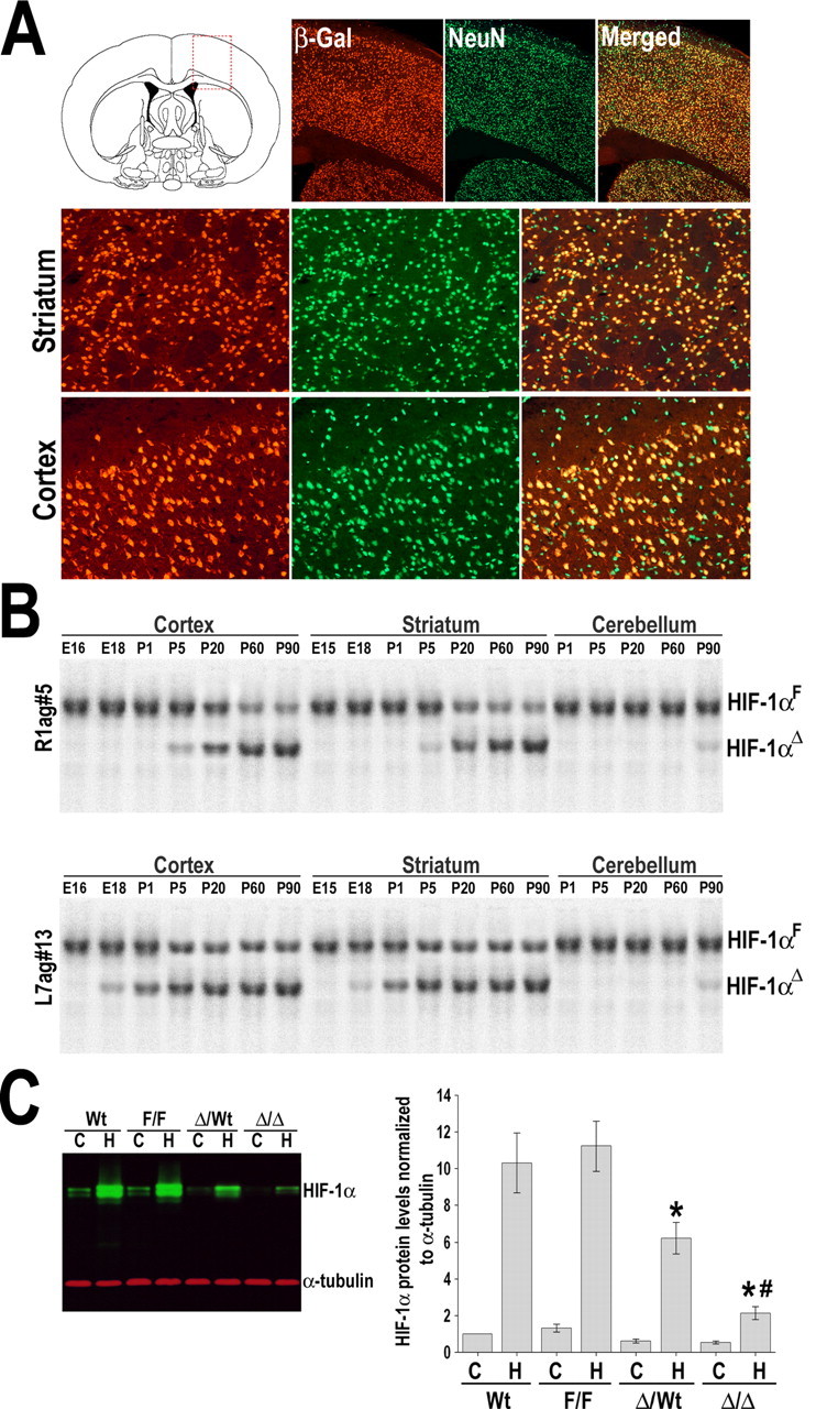
Neuron-specific knockdown of HIF-1α. A, Representative microscopic views of boxed area in diagram are shown. CaMKIIα–Cre recombinase transgenic line (R1ag#5) were mated with a LacZ reporter mouse (ROSA26Sor), and brains from offspring (P20) were analyzed by β-galactosidase staining (red). Double staining with NeuN (green) demonstrated neuronal β-galactosidase production. Bottom panels show higher-magnification views of the striatum and cortex. B, Southern blot analysis was performed on DNA isolated from the cerebral cortex, striatum, and cerebellum of Cre(+):HIF-1αF/F mice at indicated ages (E16 to P90), to detect floxed undeleted allele (HIF-1αF) and deleted allele (HIF-1αΔ). C, Decreased cerebral HIF-1α expression at normoxic (C) and hypoxic (H) (8% O2 for 4 h) conditions was confirmed in R1-HIF-1αΔ/Δ mice by Western blot analysis. Heterozygous HIF-1αΔ/wt also showed a moderate reduction of hypoxic HIF-1α levels. Data are expressed as the mean ± SD from four different experiments. *p ≤ 0.05 versus hypoxic wild-type (Wt-H). # p ≤ 0.05 versus hypoxic heterozygotes HIF-1αΔ/wt.