


Figure 12.
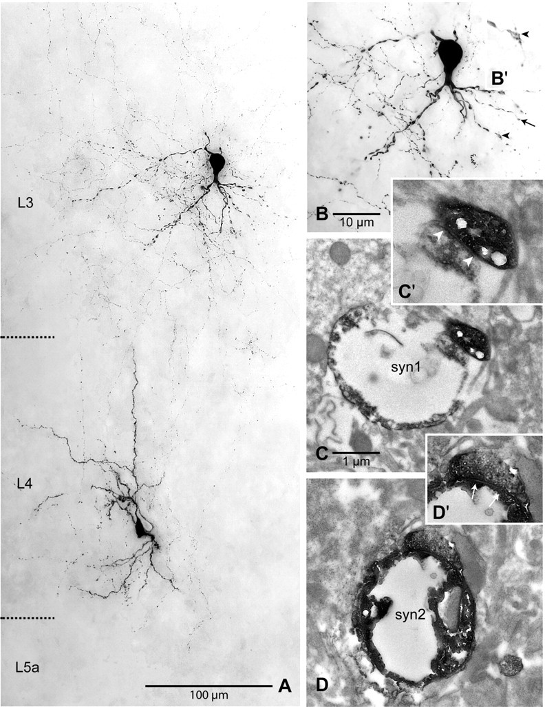
Correlated light- and electron-microscopic analysis of a synaptically coupled L4 spiny stellate neuron–layer 2/3 interneuron pair. A, Light-microscopic maximum intensity projection showing the somata, dendrites, and a part of the axons of two biocytin-filled neurons. The maximum intensity projection covered ∼60 μm in thickness of the slice. Note the numerous ascending axonal collaterals of the spiny stellate neuron. Cortical layers are indicated on the left. B, Light-microscopic maximum intensity projection of the L2/3 interneuron at higher magnification showing the varicose dendritic tree, most of which is oriented toward the white matter. Two separate very thin axonal branches originating from the spiny stellate neuron contact a secondary (arrow) and a tertiary (arrowhead) dendrite, forming putative synapses. C, Electron-microscopic verification of an asymmetric synapse (“syn1”) formed by an axonal bouton with the varicose dendritic segment of the tertiary dendrite. In C′, the synaptic cleft (arrowheads) can be seen more clearly at higher magnification. D, Also the second light microscopically observed contact was verified as an asymmetric synapse (“syn2”) on electron microscopic examination. In D′, the synaptic cleft (arrows) can be seen more clearly at higher magnification. Scale bars: A, 100 μm; B, 25 μm; C, D, 1 μm.