


Figure 1.
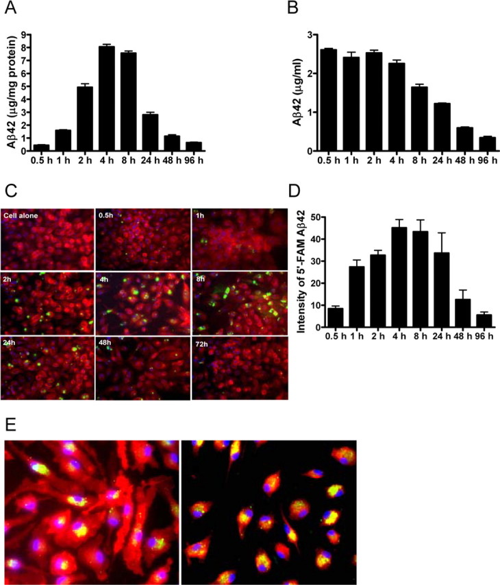
Macrophages degrade exogenous Aβ42. Human Aβ42 (3 μg/ml) was incubated with macrophages for the indicated times. A–C, Aβ42 levels in the cell pellet (A) or in the media (B) after incubation were measured by ELISA. FAM-Aβ42 (1 μg/ml; green) was incubated with macrophages (labeled red with F4/80) for the indicated times (C). Cell nuclei were visualized by DAPI (blue). Original magnification, 40×. D, The intensity of intracellular FAM-Aβ42 was quantified using Image-Pro Plus software. E, Colocalization (yellow) of FAM-Aβ42 (green) with macrophages (red; left panel) or the endosomal/lysosomal marker LAMP-1 (red; right panel). Macrophages were incubated with 1 μg/ml FAM-Aβ42 for 2 h and imaged with fluorescence microscopy. Cell nuclei were visualized with DAPI (blue). Original magnification, 40×. Representative data from one experiment that was repeated three times with similar results. Error bars represent the SE measurement.