


Figure 4.
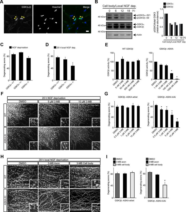
GSK3β, and not GSK3α, is a key regulator axon degeneration in vitro. A, Cultured DRG immunocytochemistry shows GSK3 enrichment in the soma. The yellow arrowheads point to neuronal cell bodies. B, Phosphorylation state of GSK3 isoforms in the cell body compartment following distal axon NGF deprivation. Over a period of 18 h, both isoforms of GSK3 are dephosphorylated, indicating elevated kinase activity. Representative blot from three independent experiments is shown. Relative abundance of pGSK3α/total GSK3α and pGSK3β/total GSK3β are shown in the adjacent graph. Note the difference in total protein loaded for each lane. C, E13.5 DRG explants from GSK3α+/+, GSK3α+/−, and GSK3α−/− embryos were cultured overnight, and then deprived of NGF for 20 h. Genetic deletion of GSK3α has no effect on axon degeneration following NGF deprivation. Values represent mean ± SEM (n ≥ 3). D, Dissociated DRG neurons from GSK3α+/+, GSK3α+/−, and GSK3α−/− were cultured for 5 d in Campenot chambers and then deprived of NGF for 28 h. Genetic deletion of GSK3α does not significantly reduce local axon degeneration. Values represent mean ± SEM (n ≥ 5). E, HEK293 cells were transfected with wild-type GSK3β or GSK3β V110I/L132A/F175L. Kinase activities were determined in the presence of varying concentrations of 3MB-PP1 using anti-V5 immunoprecipitates and a continuous fluorescent kinase assay. 3MB-PP1 potently inhibits GSK3β-ASKA activity but not wild-type activity. Values represent mean ± SEM (*p < 0.05, Wilcoxon's test, 25 μg of lysate, 20 μm ATP, n = 3). F, Images of DRG axons after 20 h NGF deprivation, cultured from ASKA mutants or wild-type animals. In the presence of ASKA inhibitor 3MB-PP1, ASKA mutants show significantly less degeneration. Wild-type explants degenerate with 3MB-PP1, indicating that GSK3β inhibition is specific. G, Quantification of reduced axon degeneration of GSK3β-ASKA knock-in DRGs in the presence of ASKA inhibitors. Values represent mean ± SEM (*p < 0.05, Student's t test; n = 9). H, Compartment-specific inhibition of GSK3β using chemical genetics. DRGs from GSK3β-ASKA knock-in embryos were cultured in Campenot chambers for 5 d. 3MB-PP1 (10 μm) was added to either the cell body or axon compartment immediately following local NGF deprivation. 3MB-PP1 potently inhibits axon degeneration when applied to the cell body compartment alone, but not the axon compartment alone. I, Quantification of reduced local axon degeneration, specifically in GSK3β-ASKA knock-in neurons exposed to 3MB-PP1 in the cell body compartment. Values represent mean ± SEM (*p < 0.05, Student's t test; n ≥ 6). Scale bar: A, 60 μm. The high-magnification insets in F and H are 62 μm squares.