


Fig. 2.
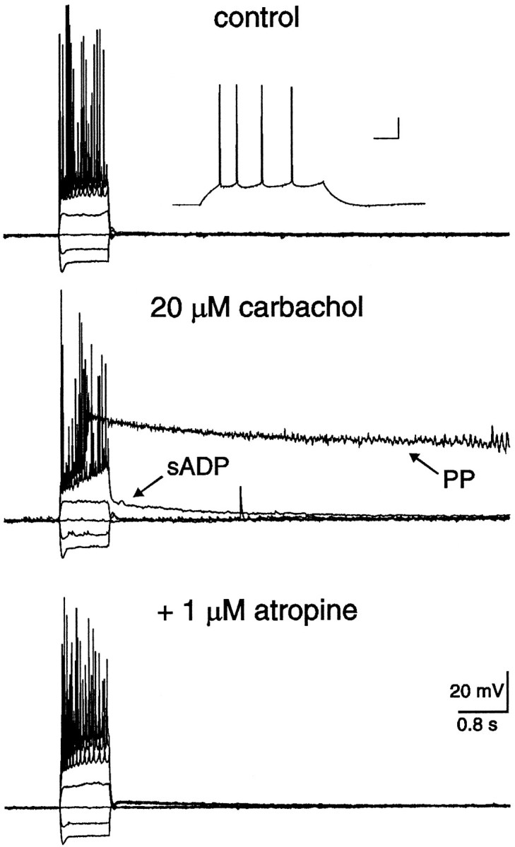
Both the sADP and PP were depressed by coapplication of the muscarinic receptor antagonist atropine. In control, the resting membrane potential of this neuron was −65 mV, and neither the sADP nor the PP was observed after action potential firing. The inset illustrates the typical firing pattern of this CA1 pyramidal neuron. The calibration bars in the insetrepresent 40 msec and 20 mV. In the presence of 20 μm carbachol, the membrane potential depolarized to −61 mV, and both the sADP and PP were observed after the spike firing. Coapplication of 1 μmatropine depressed both the sADP and PP, implicating the involvement of muscarinic receptors.