


Fig. 3.
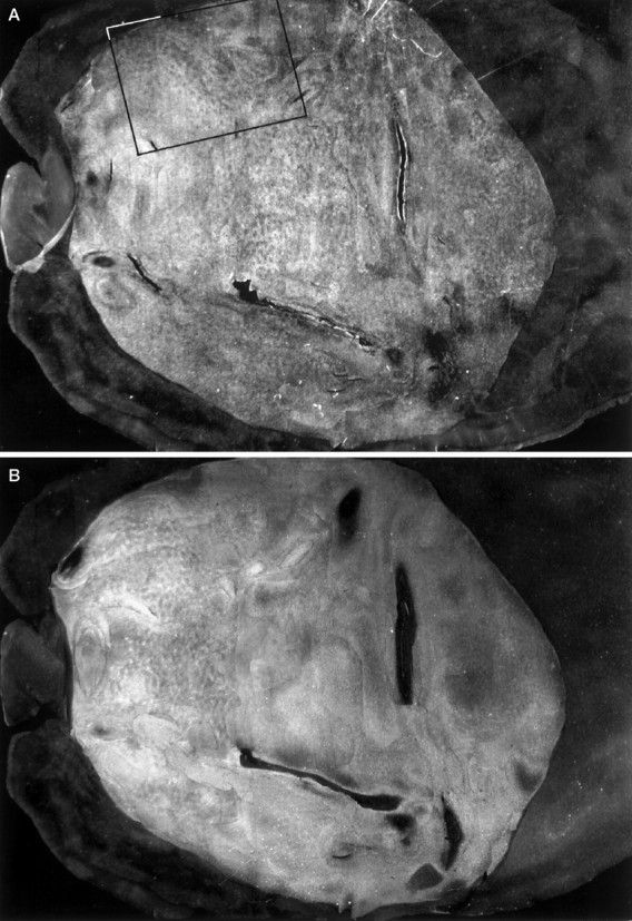
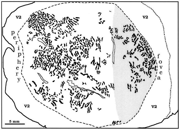
A, Montage of WGA-HRP label from the left eye in layer IVcα of the right cortex of monkey 4. Faint columns are visible throughout most of the montage. Therectangle delineates the portion of the flat-mount shown at higher power in Figure 4. B, Montage of autoradiographs through layer IVcα showing columns of [3H]proline label from the right eye. C, Sketch of the columns labeled in V1, prepared by combining the WGA-HRP data in A and the proline data in B. The contrast of the columns in B was reversed so that theblack columns throughout the figure represent the ocular dominance columns of the right eye. They form an irregular array of short stripes and islands, with a mean diameter of 225 μm. Theshaded area corresponds to the operculum, grossly visible in Figure 1, representing the central 3–4°. Figure 3continues.