


Fig. 5.
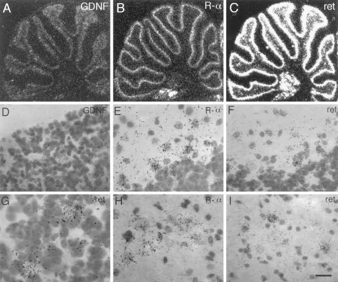
Complete trophic circuits within the cerebellum.A–C, Autoradiograms of hybridized sagittal sections through the cerebellum show that GDNF, RET, and GDNFR-α mRNAs are all expressed in the cerebellar cortex as well as the deep cerebellar nuclei. D, Bright-field photomicrograph of the granular layer reveals that scattered cells within this layer that do not seem to be granule cells express GDNF mRNA. E, GDNFR-α mRNA is seen to be expressed most highly in the Purkinje layer in cells directly adjacent to Purkinje cells (possibly basket cells) and also at lower levels reaching into the molecular layer.F, RET mRNA is also observed in cells similarly placed next to Purkinje cells, as well as within the molecular layer.G, High-magnification bright-field photomicrograph of the granular layer of the cerebellum reveals that RET mRNA is highly expressed in cells that stain very lightly with cresyl violet.H, I, Neurons of deep cerebellar nuclei are seen to express both GDNF receptors. Scale bar (shown inI): D, 29 μm; E, F, H, I, 20 μm; G, 14 μm.