


Fig. 6.
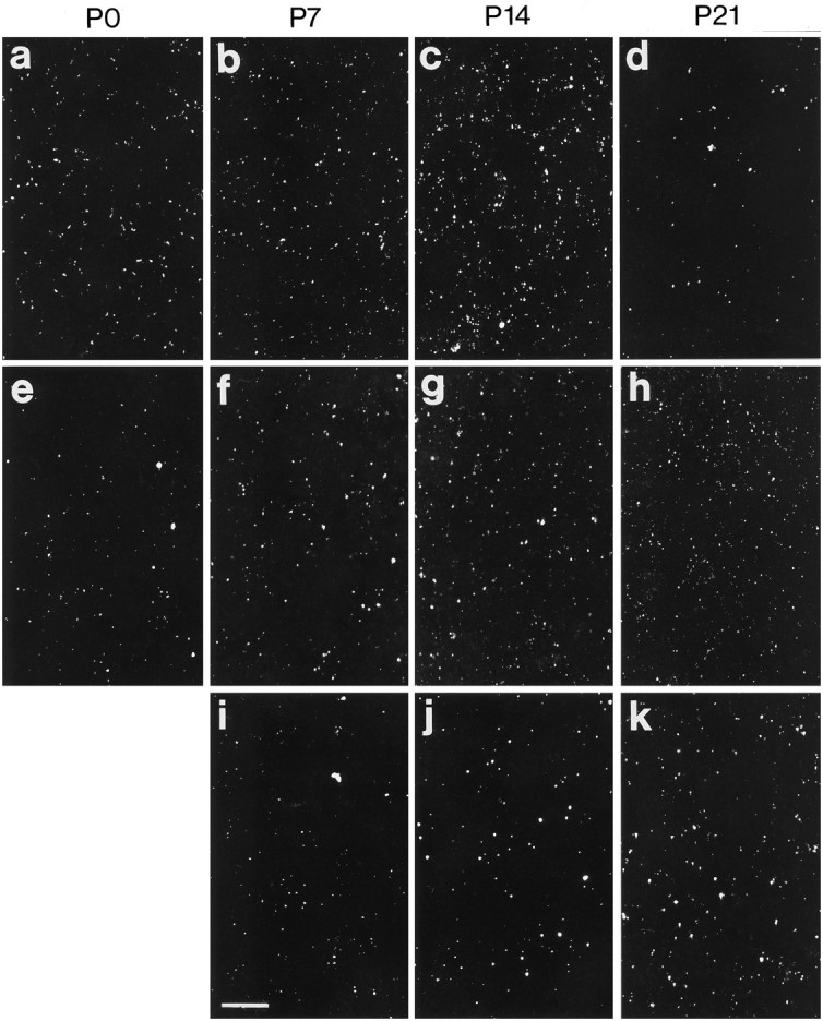
Examples of Cxs 26 (a–d), 43 (e–h), and 32 (i–k) immunolabeling in the lower layers of the visual cortex during postnatal development (P0–P21). Note the differences between the three Cxs with regard to particle density and particle size in the developing cortex. Scale bar, 40 μm.