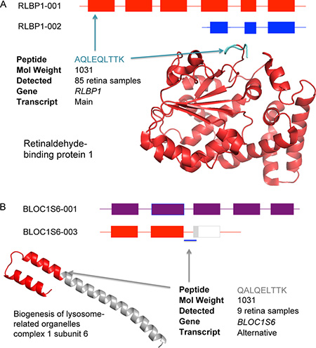
Box 3, Figure I. Identifying two peptides from the same spectrum.
(A) The peptide AQLEQLTTK is from the main isoform of RLBP1 (Retinaldehyde-binding protein 1), a protein expressed in retina. The structure of RLBP1 has been resolved and is shown bottom right; the position of peptide AQLEQLTTK is marked in blue. (B) Peptide QALQELTTK supports the presence of an alternative isoform of BLOC1S6 that would cause the loss of the large coiled coil region shown in grey in the figure.