


Figure 6.
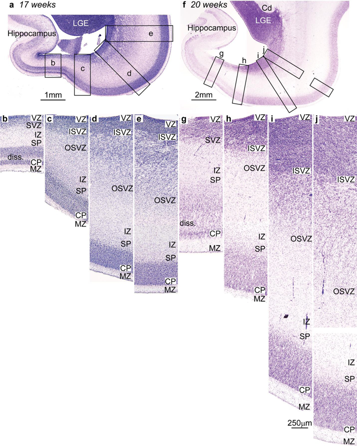
Systematic variation of embryonic layers in the prospective temporal lobe of the human embryo (Nissl staining). Germinal zones and prospective cortical layers are named according to Smart et al. (2002) and Bystron et al. (2008). a-e, Photomicrographs from a human fetus of 17 weeks gestational age stained for Nissl. a, Low power photomicrograph at the level of the temporal lobe shows the levels of photomicrographs b-e. b, The germinal zones of the prospective entorhinal cortex next to the hippocampal formation are composed of thin ventricular (VZ) and subventricular (SVZ) zones; the SVZ has no sublayers. c, The SVZ of the prospective perirhinal cortex is divided into inner (ISVZ) and outer (OSVZ) zones. d, The OSVZ is more prominent in prospective lateral (eulaminate) temporal areas. e, The cell density and the thickness of the OSVZ increases progressively in a lateral direction along a line of increasing laminar elaboration. f-j, Photomicrographs from a human fetus of 20 weeks gestational age stained for Nissl. f, Low power photomicrograph at the level of the temporal lobe shows the levels of photomicrographs g-j. g, The germinal zones of the prospective entorhinal cortex next to the hippocampal formation are thicker compared to b, but the SVZ still does not show two subzones. h, The prospective perirhinal cortex has thicker VZ and SVZ; the latter is subdivided into ISVZ and OSVZ. i, Prospective lateral eulaminate areas have thicker VZ and ISVZ than prospective entorhinal and perirhinal areas; the OSVZ predominates. j, The thickness of the OSVZ increases progressively in the lateral direction consistent with increased laminar definition in the adult. Abbreviations: Cd: caudate; CP, cortical plate; ISVZ, inner subventricular zone; IZ, intermediate zone; LGE, lateral ganglionic eminence; MZ, marginal zone; OSVZ, outer subventricular zone; SP, subplate; SVZ, subventricular zone; VZ, ventricular zone. Calibration bar in i applies to b-e and g-j. [Note: This figure is a re-examination of material from an earlier paper (Reillo et al. 2011)].