


Figure 5.
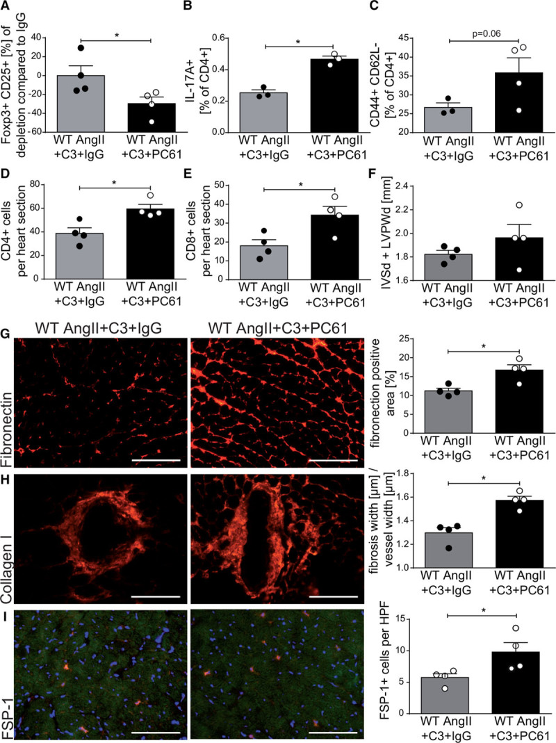
Depletion of regulatory T cells abrogates the effect of propionate in AngII-infused wild-type NMRI (WT) mice. AngII-infused WT mice received propionate treatment with intraperitoneal injections of anti-CD25 (PC61) or IgG control. A, Relative reduction in splenic CD4+CD25+Foxp3+ regulatory T cells at day 14 of AngII infusion in comparison with IgG control. B, IL-17A production in CD4+ restimulated splenocytes measured by flow cytometry. C, Splenic effector memory T cell (CD4+CD44+CD62L–) frequencies. D and E, Analysis of CD4+ (D) and CD8+ (E) lymphocytes in heart sections using immunofluorescence. F, Left ventricular wall thickness (sum of IVSd and LVPWd) as measured by echocardiography. G through I, Immunofluorescence analysis of left ventricular fibrosis using fibronectin (G), collagen I (H), and FSP-1 (I) antibodies. Left, Representative photomicrographs (scale bars=100 µm). Right, quantification. A, D through I, WT AngII+C3+IgG n=4, WT AngII+C3+PC61 n=4; B and C, WT AngII+C3+IgG n=3, WT AngII+C3+PC61 n=4, *P<0.05 by 1-tailed Mann-Whitney test. AngII indicates angiotensin II; C3, propionate; FSP-1, fibroblast-specific protein 1; IgG, immunoglobulin G; IL, interleukin; IVSd, interventricular septal thickness at diastole; and LVPWd, left ventricular posterior wall end diastole.