


Figure 3.
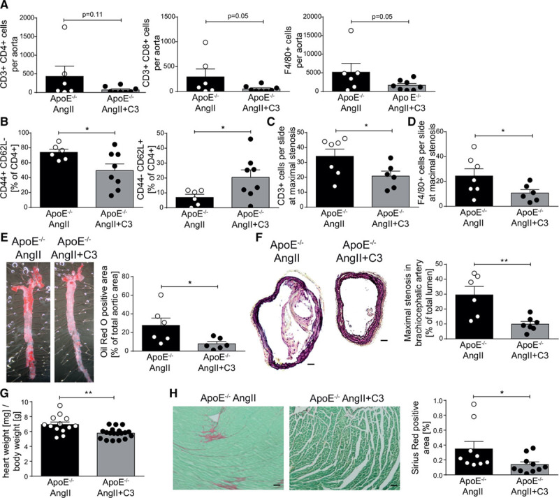
Propionate reduces aortic inflammation and atherosclerotic lesion burden in AngII-infused ApoE–/–. A,Single-cell suspensions from whole aortas of were analyzed for T helper (CD3+CD4+), cytotoxic T cells (CD3+CD8+), and macrophages (F4/80+) by flow cytometry. B, Aortic CD4+ T cells were analyzed for CD4+ effector memory (CD44+CD62L–) and naive (CD44–CD62L+) subsets by flow cytometry. C and D, Quantification of CD3 and F4/80 positive cells in sections of the brachiocephalic artery, respectively. E, En face Oil Red O staining of whole aortas for the quantification of atherosclerotic lesion burden. Left, Representative aortas. Right, Quantification. F, The degree of stenosis in the brachiocephalic artery was determined in Movat-stained cross-sections. Left, Representative sections (scale bar=100 µm). Right, Quantification. Athrough F, ApoE–/– AngII n=6, ApoE–/– AngII+C3 n=8. G, Cardiac hypertrophy index (heart weight [mg]/body weight [g]) of AngII-infused ApoE–/– mice treated with C3 or control, ApoE–/– AngII n=16, ApoE–/– AngII+C3 n=21. H, Left ventricular cardiac fibrosis as analyzed by Sirius red staining. Left, Representative photomicrographs (scale bar=100 µm). Right, Quantification. ApoE–/– AngII n=9, ApoE–/– AngII+C3 n=10. *P<0.05, **P<0.01 by 1-tailed t test or Mann-Whitney test. AngII indicates angiotensin II; ApoE–/–, apolipoprotein E knockout–deficient; and C3, propionate.