


FIGURE 2: Lineage tracing using Tie2-CreERT2;R26R in Col1a2oim/oim or Fbn1C1039G/+ mutant backgrounds as an indicator of EMT during disease.
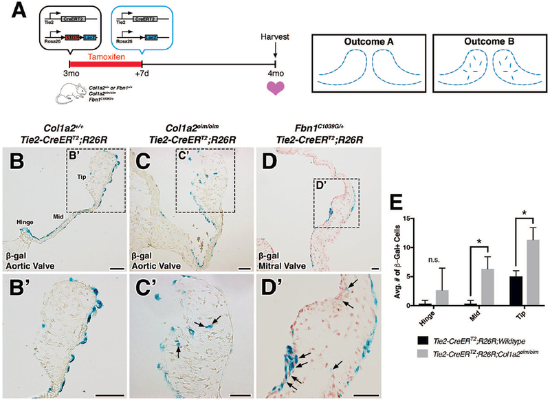
A: Schematic of experiment. Tamoxifen chow was administered to 3 month-old Col1a2+/+;Fbn1+/+ or mutant transgenic mice for 7 days. Cre-mediated recombination results in the deletion of stop codons and expression of β-galactosidase (β-gal) from the lacZ gene. At 4 months-of-age, hearts were harvested, sectioned, and analyzed for β-gal expression. In the absence of EMT, β-gal+ cells will be located solely on the VECs lining the valves (outcome A). In the presence of an EMT event, β-gal+ cells will be detected within the valve interstitium (outcome B). B-D: Histologic sections of aortic (B, B’, C, C’) and mitral (D, D’) valves from β-gal stained wildtype, Col1a2oim/oim, and Fbn1C1039G/+ transgenic mice. In mutant transgenic mice, β-gal+ cells were detected within the valve interstitium (arrows). E: Quantification of β-gal+ cells within the valve interstitium from Col1a2oim/oim and Col1a2+/+ wildtype controls. Student’s t-test was used with statistical significance defined as *p<0.05. Data are reported as mean, and error bars represent standard deviation. Scale Bar = 50μm.