


Figure 4. Preservation of Proteins and Structure of CC in Cep290rd16/rd16;Nrl—/— Mice by AAV8-Myo-tail Vector.
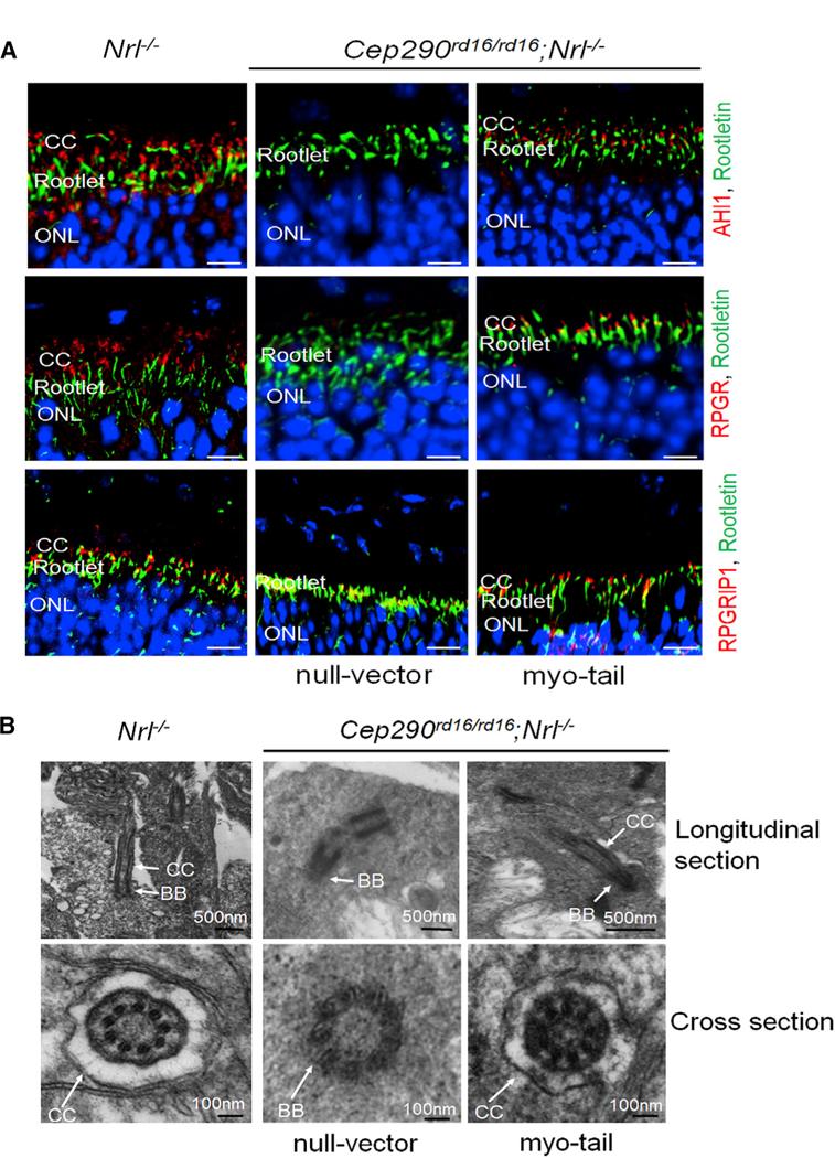
Each mouse was injected with 1 × 10⁹ vg AAV8-myo-tail vector in one eye and the same dose of a null vector in the fellow eye at P14.
(A) Representative confocal images of retinal sections from 4.5-month-old Cep290rd16/rd16;Nrl—/— mice showing the preservation of AHL1, RPGR, and RPGRIP1 at CC following myo-tail vector treatment. Sections of an age-matched Nrl—/— mouse were used as controls. Scale bars, 20 µm.
(B) Representative images of transmission electron microscopy showing relatively normal connecting cilium structure with myo-tail vector treatment. No connecting cilium structure was found in eyes treated with the null vector.
BB, basal body; CC, connecting cilia.