


FIG. 1.
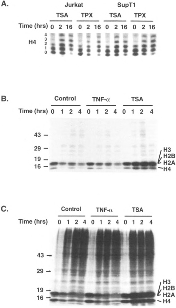
TSA and TPX inhibit histone deacetylation in lymphoid cell lines. (A) Histones were acid extracted from Jurkat or SupT1 cells treated with TSA (400 nM) or TPX (10 nM) for 0, 2, or 16 h and separated on a 15% Triton-acid-urea gel. Histones were visualized after silver staining and the portion of the gel corresponding to different acetylated isoforms of histone H4 is shown. (B, C) Jurkat cells were pulsed for 30 min with [3H]acetate (250 μCi/ml NEN), washed, and chased for 0–4 h in the presence of TNF-α (80 U/ml) or TSA (400 nM). Nuclei were either acid extracted and the purified histones loaded on a 12% polyacrylamide denaturing gel (B) or directly resuspended in Laemmli buffer and loaded on a 12% polyacrylamide denaturing gel to examine all nuclear proteins (C). Molecular weight markers in kDa are indicated on the left of the gel. The position of different histones is indicated on the right.