


Figure 2. Bre2/ASH2L fold into an extended non-canonical SPRY domain.
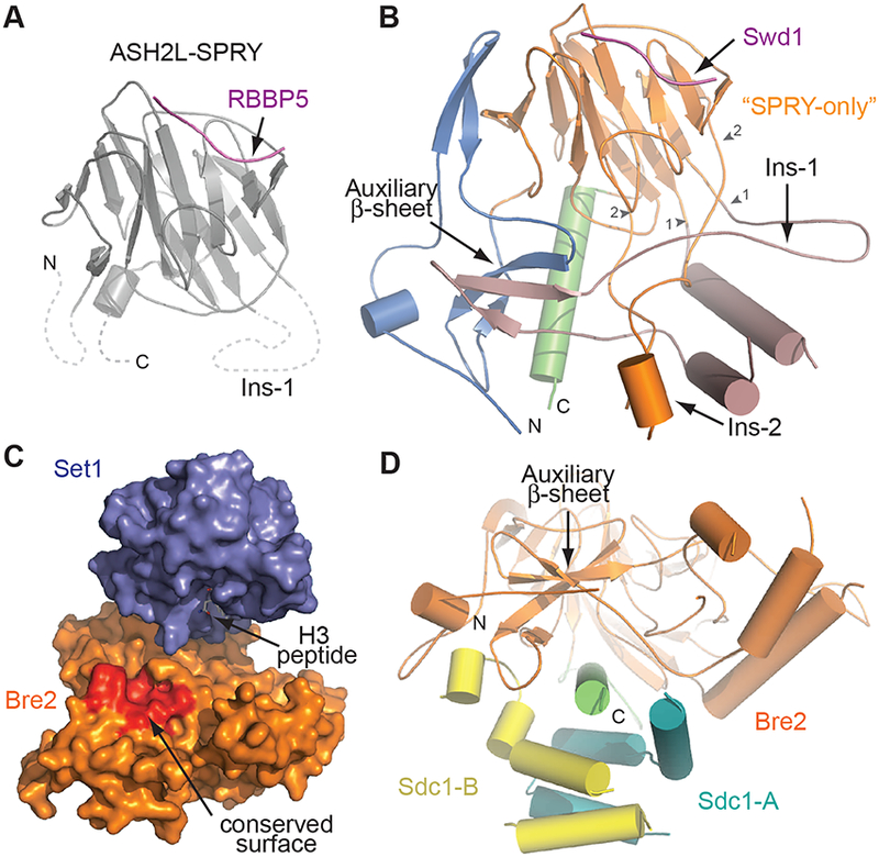
(A) Crystal structure of human ASH2L (grey) bound to a peptide of RBBP5 (magenta) (PDB: 4X8P). Dashed lines indicate the absent N-, insertion, and C-terminal sequences in the structure.
(B) Structure of yeast Bre2. The conserved SPRY-only domain and Ins-2 is shown in orange, while the N-terminal pre-SPRY and Ins-1 are shown in blue and pale pink, respectively. The C-terminal helix is colored in green. Small arrowheads indicate the terminal ends of the insertion elements. The Swd1 peptide equivalent to that of RBBP5 in (A) is shown in purple. Structure is depicted in smooth lines representation for clarity.
(C) Surface representation of Set1 (blue) and Bre2 (orange). A highly conserved surface formed by the auxiliary β-sheet in Bre2 is highlighted in red.
(D) View of Bre2 looking down the C-terminal helix (green), illustrating the position of the auxiliary β-sheet right above the helix. Sdc1-A (yellow) and Sdc1-B (turquoise) prop up this sheet.