


Fig. 2. in situ vaccination of CpG in combination with anti-OX40 antibody cures established local and distant tumors.
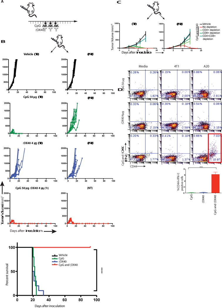
(A) Treatment schema. BALB/c mice were implanted subcutaneously with A20 lymphoma cells (5 × 10⁶) on both the right and left sides of the abdomen. When tumors reached between 0.5- 0.7cm in the largest diameter (typically on day 4-5 post inoculation), αOX40 (4μg) and CpG (50μg) were injected into one tumor site every other day for a total of three doses. Tumors sizes were serially measured by caliper. (B) Tumor growth curves. Left column: treated tumors (Tr), right column: non-treated tumors (NT). Top to bottom: Vehicle, CpG, αX40, CpG and αOX40. Survival plots of the treated mice, (n = 10 mice per group). Shown is one representative experiment out of 9. (C) Effect of CD4/CD8 depletion. Mice were implanted with bilateral tumors and one tumor was injected with CpG and αOX40 antibody according to the schema in A. CD4 (0.5 mg) or CD8 (0.1 mg) depleting antibodies were injected i.p. on days 6, 8, 12, and 15 (n = 10 mice per group). (D) CD8 T cell immune response. Splenocytes from the indicated groups obtained on day 7 post-treatment were co-cultured with either media or with 1 × 10⁶ irradiated 4T1 (unrelated control tumor) or with A20 (homologous tumor) cells for 24h. Intracellular IFN-γ was measured in CD8+ T cells by flow cytometry as a percentage of CD44hi (memory CD8) T cells shown in dot plots and in bar graph summarizing data from three experiments (n = 9 mice per group).