


Figure 4. Arc extracellular vesicles mediate intercellular transfer of protein and mRNA in HEK293 cells.
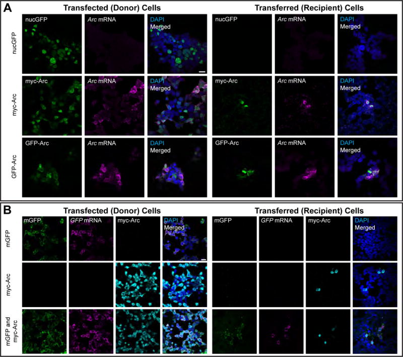
(A) Donor HEK cells in 10-cm dishes were transfected with GFP-Arc, myc-Arc, or nuclear GFP (nucGFP) for 6h. Culture media containing plasmid DNA and transfection reagents was then removed and replaced with fresh culture media. 18h later, this media was removed and used to replace media on naïve recipient HEK cells on coverslips in 12-well plates. 24h later, these cells were fixed and combined FISH for Arc mRNA and ICC for Arc protein was performed. (left) Representative images of HEK cells grown on coverslips and transfected with the same protocol as in 10-cm dishes, showing Arc protein (ICC) and Arc mRNA (FISH). (Right) Representative images of recipient HEK cells showing Arc mRNA and protein were present in cells that received media from GFP-Arc- and myc-Arc-transfected cells, but not nucGFP-transfected cells. Scale bar=20μm. Representative of 7 independent experiments and cultures. (B) Donor HEK cells in 10-cm dishes were transfected as in (A) with membrane GFP (mGFP), myc-Arc, or both constructs together. The media was replaced after 6h, and 18h later, transferred to naïve recipient HEK cells in 12-well plates. 24h later, cells were fixed and combined FISH/ICC for GFP mRNA and Arc protein was performed. (left) Representative images of transfected HEK cells grown on coverslips, showing mGFP fluorescence, Arc protein and GFP mRNA. (right) Representative images of recipient HEK cells that show co-transfer of GFP protein and mRNA with Arc protein. No GFP transfer was observed in the mGFP only group. Scale bar=20μm. Representative of 3 independent experiments and cultures.