


Figure 3. Structure of the orthosteric mAChR ligand binding site.
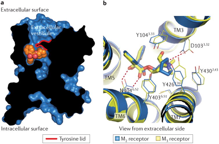
a | A cross-section through the M2 receptor structure reveals a large, solvent-accessible cavity extending through the receptor. The QNB ligand (orange spheres) is bound within the receptor transmembrane core (black, receptor protein; blue, receptor surface). b | The orthosteric ligand (antagonist)-binding sites for the M2 and M3 receptors are almost identical in both structure and sequence. Polar contacts (red dotted lines) between the receptor and bound antagonist (M2, QNB; M3, tiotropium) are identical for the two receptors. Residues are numbered according to the human M2 receptor sequence. mAChR, muscarinic acetylcholine receptor.