


Fig. 1.
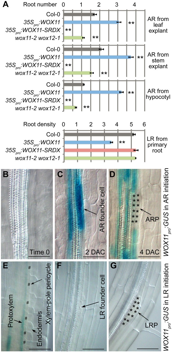
Analysis of WOX11 in AR and LR formation on medium. (A) Statistical analysis of root number per leaf explant at 12 DAC, per stem explant at 12 DAC, per hypocotyl and per 1-cm primary root. LR analysis includes emerged LRs together with LRP observed under DIC microscopy. Error bars represent s.e.m. from three biological repeats. n=30 (leaf explant, stem explant and hypocotyl) or n=10 (primary root) in each repeat. **P<0.01 (two-sample Student's t-test compared with Col-0). (B-D) GUS staining of WOX11pro:GUS from leaf explant at time 0 (B), 2 DAC (C) and 4 DAC (D). Note that the GUS signal was detected specifically in the AR founder cells (C) (Liu et al., 2014). We observed more than 20 leaf explants from two independent lines and all of them showed GUS staining in AR founder cells. (E-G) GUS staining of WOX11pro:GUS in the primary root. Note that GUS signal was detected in the protoxylem at the meristematic region (E) as previously reported (Liu et al., 2014), but was not detected in xylem-pole pericycle cells (E), LR founder cells (F) or LRP (G). We observed more than 20 seedlings from two independent lines and none of them showed GUS staining in LR founder cells. Also see Fig. S4D-F. Leaf explants (A-D) and stem explants (A) were detached from 12- and 24-day-old seedlings grown on 1/2 MS medium, respectively. The detached explants were then cultured on B5 medium without added hormones (Chen et al., 2014). Hypocotyls (A) were analyzed using 14-day-old seedlings vertically grown on 1/2 MS medium. Primary and lateral roots were analyzed using 9-day-old (A) or 7-day-old (E-G) seedlings vertically grown on 1/2 MS medium. Asterisks in D and G indicate root primordium cells, and hash symbols in E indicate xylem-pole pericycle. Scale bars: 50 μm.