


Figure 5.
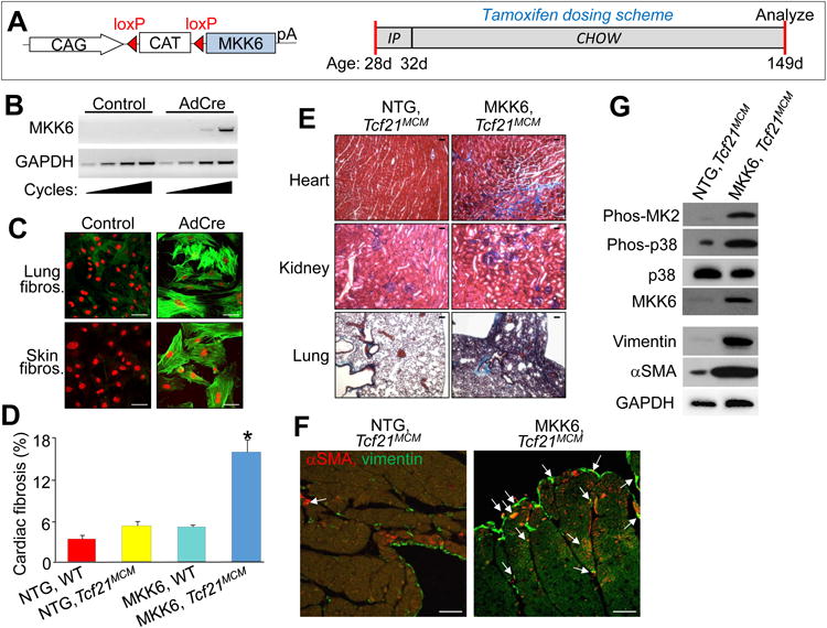
Constitutive activation of p38 in cardiac fibroblasts enhances fibrotic and hypertrophic remodeling. A, Left panel: schematic of the MKK6 LoxP-dependent and inducible transgene driven by a ubiquitous promoter (CMV β-actin). Right panel: schematic of the tamoxifen dosing scheme used for these experiments with age listed in days (d). B, RT-PCR for the activated MKK6 transgene from cardiac fibroblasts isolated from MKK6-Tg, Tcf21MCM mice and subjected to infection with Adβgal or AdCre. GAPDH expression was used for normalization. C, Immunofluorescent images of fibroblasts isolated from lung and skin of MKK6-Tg mice that were then adenovirally transduced with Adβgal (control) or AdCre and immunostained for αSMA+ stress fibers (green) and nuclei (red). Scale bar = 50 μm. D, Quantification of the area of fibrosis (blue) in transverse histological heart sections with Masson's trichrome staining in the indicated genotypes of mice. Average fibrotic area ± SEM, N≥8, *P<0.05 vs NTG and Tcf21MCM. E, Masson's trichrome-stained histological sections from the indicated tissues of either non-Tg (NTG) or MKK6-Tg mice with the Tcf21MCM allele after 4 months of continuous tamoxifen treatment. Blue staining indicates fibrosis. Scale bar = 25 μm. F, Immunofluorescent staining of hearts from the indicated genotypes of mice after 4 months of continuous tamoxifen treatment. Myofibroblasts are positive for both αSMA (red) and the fibroblast marker vimentin (green) are designated with the white arrows. Scale bar = 100 μm. G, Western blot analysis for the indicated proteins from cardiac fibroblasts purified from Tcf21MCM and MKK6-Tg, Tcf21MCM hearts following 1 month of tamoxifen induction. GAPDH antibody was used as a loading control.