


Figure 2.
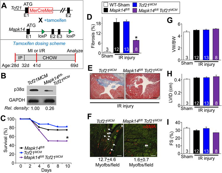
Mapk14 deletion in resident cardiac fibroblasts minimizes the fibrotic response to ischemic injury. A, Top panel: schematic showing the Tcf21 locus containing a tamoxifen-regulated MerCreMer (MCM) cDNA and a Mapk14-loxP targeted allele contained within mice so that with tamoxifen administration Cre-dependent recombination can inactivate the Mapk14 gene. Lower panel: schematic of the tamoxifen dosing scheme used for these experiments with age listed in days (d). B, Western blot analysis for p38α protein from purified cardiac fibroblasts isolated from hearts of Mapk14fl/fl Tcf21MCM mice after tamoxifen induction. GAPDH was used as a loading control. C, Kaplan-Meier plot of survival of the indicated genotypes of mice over 10 days after IR injury. D, Quantification and E, Masson's trichrome stained histological images from the hearts of the indicated mice 28 days after IR injury. Blue staining indicates fibrosis. Scale bar = 150 μm. Average fibrotic area ± SEM, N=3 for sham and N≥8, *P<0.05 vs Mapk14fl/fl and Tcf21MCM. F, Immunofluorescent staining and quantification of myofibroblast numbers (white arrows) in the cardiac infarction border zone 28 days after injury. Myofibroblasts are αSMA (red) positive and negative for isolectin-B4 (green). Scale bar = 50 μm. Data represent the average number of αSMA+/isolectin-B4- cells/section ± SEM, 5 sections were measured per heart, N=3 for sham and N≥8 mice per group. G, Quantification of ventricular weight (VW) normalized to body weight (BW) 28 days after ischemic injury, Average VW/BW ± SEM, *P<0.05 vs Mapk14fl/fl and Tcf21MCM. Echocardiography of H, left ventricle inner diameter (LVID) and I, fractional shortening (FS%) from Mapk14fl/fl-Tcf21MCM, Mapk14fl/fl and Tcf21MCM mice 28 days post IR injury. Data are averages ± SEM, *P<0.05 vs Mapk14fl/fl and Tcf21MCM. The legend above panels D and E represent all of the data in the figure. Number of mice analyzed is shown within the bars of panels D,G-I