


Figure 3.
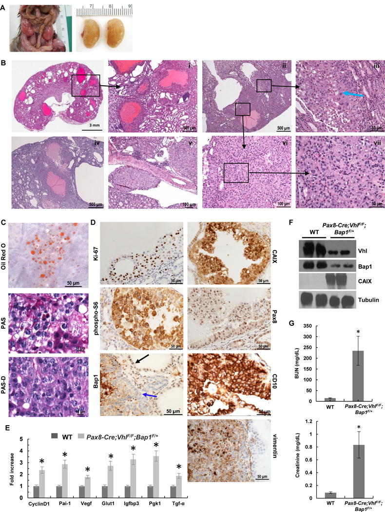
Pax8-Cre;VhlF/F;Bap1F/+ mice develop high grade ccRCC. A, Representative images from Pax8-Cre;VhlF/F;Bap1F/+ mutant mice showing enlarged nodular kidneys. B, H&E staining of kidney sections from Pax8-Cre;VhlF/F;Bap1F/+ mice (n=14) showing (i) cystic and solid ccRCCs, (i,iv) multiple large cystic tumors with marked proliferative lining, (iii,vi,vii) solid tumor with thin arborizing vascular network, (iii,vii) neoplastic cells with marked pleomorphism, anaplasia, prominent nucleoli, and mitosis (blue arrow indicates atypical mitosis), and (v) lymphovascular spread. C. Oil Red O staining for triglycerides and neutral lipids, and Periodic acid-Schiff staining without (PAS) and with (PAS-D) diastase highlight the abundant lipid and glycogen in neoplastic cells. D, IHC for Ki-67, CAIX, phospho-S6, Pax8, Bap1 (blue arrow, tumor cells; black arrow, retained Bap1 staining), CD10, and vimentin in RCCs. Complete loss of nuclear Bap1 expression in Pax8-Cre;VhlF/F;Bap1F/+ mice was seen in 30% (21/69 lesions (n=8)), however, no appreciable differences in morphology, size, or grade across lesions were seen. E, qRT–PCR for the indicated Hif target genes in kidneys of Pax8-Cre;VhlF/F;Bap1F/+ mice and controls (n=3 independent mice; PCR done in triplicate). F, Western blot analysis of the indicated proteins in Pax8-Cre;VhlF/F;Bap1F/+ mice and controls. G, Plasma BUN and Creatinine measurements in Pax8-Cre;VhlF/F;Bap1F/+ mice and controls (n=3 independent mice for each group). *, P < 0.05. (WT, age-matched VhlF/F;Bap1F/+ mice without Cre.)