


Figure 1. LacZ expression in Tbx18:nlacZ knock-in mice recapitulates endogenousTbx18 expression.
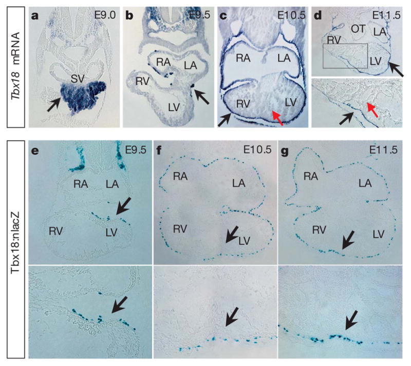
a–d, Section RNA in situ hybridization (ISH) of Tbx18 in mouse embryos (E9.0–E11.5). Tbx18 is expressed very early in the proepicardium (a) and epicardium (b–d). For parallel whole-mount RNA ISH, see Supplementary Fig. 2. e–g, X-gal staining on cardiac sections from Tbx18:nlacZ embryos at E9.5–E11.5. Tbx18:nlacZ cells are detected in the early epicardium at E9.5 (e) and in all epicardial cells covering the heart after E10.5 (f, g, data not shown for E12.5–E13.5). X-gal staining on Tbx18:nlacZ mouse tissues is consistent with Tbx18 mRNA ISH. For the Tbx18:nlacZ targeting strategy, see Supplementary Fig. 3. Black arrows indicate Tbx18 mRNA expression (a–d) and Tbx18:nlacZ cells (e–g); red arrows in c and d indicate that Tbx18 is not expressed in heart between E9.5 and E11.5. For e–g, lower panels are high-magnification views of the upper panels in the heart (black arrow regions). LA/RA, left/right atrium; LV/RV, left/right ventricle; OT, outflow tract; and SV, sinus venosus.