



Abstract
Adult neurogenesis, a developmental process of generating functionally integrated neurons, occurs throughout life in the hippocampus of the mammalian brain and showcases the highly plastic nature of the mature central nervous system. Significant progress has been made in recent years to decipher how adult neurogenesis contributes to brain functions. Here we review recent findings that inform our understanding of adult hippocampal neurogenesis processes and special properties of adult-born neurons. We further discuss potential roles of adult-born neurons at the circuitry and behavioral levels in cognitive and affective functions and how their dysfunction may contribute to various brain disorders. We end by considering a general model proposing that adult neurogenesis is not a cell-replacement mechanism, but instead maintains a plastic hippocampal neuronal circuit via the continuous addition of immature, new neurons with unique properties and structural plasticity of mature neurons induced by new-neuron integration.
Keywords: neural stem cells, development, brain disorders, dentate gyrus
INTRODUCTION
One major advance in modern neuroscience is the appreciation of the extent of plasticity in the mature nervous system. Many critical functions of the nervous system depend on its mutability: its ability to perceive external information, to encode novel associations among events and objects in the world, and to generate adaptive behavior. Plasticity thought to underlie these functions occurs at multiple levels, from epigenetic modifications of gene expression, to neuronal activity-dependent modulation of synaptic strength, to tuning of integrated circuits that carry multimodal sensory information. Perhaps the most striking form of structural plasticity in the adult nervous system is the de novo generation and integration of new neurons into the existing circuitry through a process known as adult neurogenesis (Kempermann & Gage 1999). Originally thought to occur only during embryonic development, active adult neurogenesis has now been shown in almost all mammalian species examined so far (Lledo et al. 2006, Ming & Song 2005). Active neurogenesis occurs in two discrete regions: the subventricular zone of the lateral ventricle, from where newborn neurons migrate to the olfactory bulb or striatum (Ernst et al. 2014) and differentiate mostly into interneurons, and the dentate gyrus of the hippocampus, where newborn granule cells are integrated into the local circuitry (Ming & Song 2011) (Figure 1a). Significant progress has been made in the past decade to understand the generation, development, and integration of adult-born neurons, molecular and regulatory mechanisms, and potential contributions of adult neurogenesis to brain function and dysfunction (Ming & Song 2011, Zhao et al. 2008).
Figure 1.
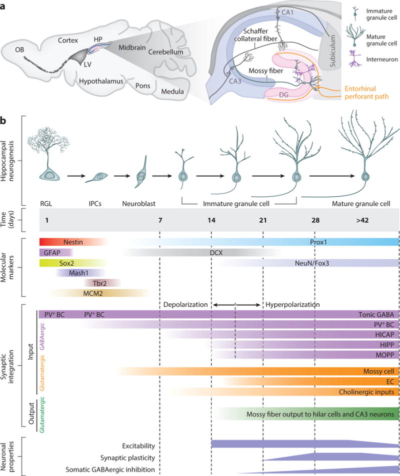
Summary of basic processes of neurogenesis in the young adult mouse hippocampus. (a) A sagittal section view of an adult rodent brain highlighting two restricted regions that exhibit active adult neurogenesis—the hippocampus (HP) and lateral ventricle (LV)—which generate new neurons that mostly migrate into the olfactory bulb. More detailed hippocampal structure is further illustrated with the primary trisynaptic circuit formed by three principal neuronal subtypes. (b) Summary of the developmental processes of adult hippocampal neurogenesis, including time course of marker expression, developmental stages, synaptic integration, and special neuronal properties associated with different stages. BC, basket cells; DG, dentate gyrus; EC, entorhinal cortex; HICAP, hilar interneuron with commissural-associational pathway-associated axon terminals; HIPP, hilar perforant path-associated interneurons; IPCs, intermediate progenitor cells; MOPP, molecular-layer perforant pathway cells; OB, olfactory bulb; PV+, parvalbumin-expressing interneurons; RGL, radial glia-like cell.
Adult hippocampal neurogenesis has garnered significant interest because of its potential to influence information processing in the medial temporal lobe, a neural substrate for many forms of learning and memory and a site of pathophysiology associated with various neurological disorders (Squire 1992). The canonical signaling network of the hippocampus consists of synaptically connected principal neurons located in three major subregions to form the trisynaptic circuit: granule cells in the dentate gyrus and pyramidal neurons in CA1 and CA3 (Figure 1a). Information flows from the entorhinal cortex through medial and lateral perforant pathways to the dentate gyrus, then to CA3 pyramidal cells via mossy fiber axons of granule cells, then to CA1 pyramidal cells via Schaffer collateral projections of CA3 neurons, and finally to the subiculum and back to the entorhinal cortex. This primary hippocampal circuit forms a closed loop wherein sensory information from specific cortical areas converges onto the entorhinal cortex, is processed through the hippocampal circuitry, and returns to the region of origin in the entorhinal cortex. In addition, there are direct projections from the entorhinal cortex to CA3 and CA1 and multiple modulatory inputs from other brain regions to the hippocampus. The dentate gyrus also contains diverse γ-aminobutyric acid (GABA)-ergic inhibitory neurons within the hilus region and the molecular layer, which mediate feedforward and feedback inhibition, and hilar glutamatergic mossy cells, which receive inputs primarily from granule cells and then innervate granule cells and local interneurons.
Within the hippocampus, dentate granule cells are the only neurons to be continuously generated. Young adult rats generate an estimated 9,000 new cells each day in the dentate gyrus, about 6% of the total granule cell population each month (Cameron & McKay 2001), whereas adult humans add 700 new neurons in each hippocampus per day, corresponding to an annual turnover of 1.75% of the renewing neuronal population (Spalding et al. 2013). The significant number of new neurons, together with dynamic regulation of adult neurogenesis by various physiological and pathological stimuli (Ma et al. 2009), suggests that adult neurogenesis may be integral to certain brain functions. Indeed, behavior analyses in animal models support a critical role for dentate newborn neurons in several hippocampus-dependent functions (Aimone et al. 2011). Electrophysiological analyses have identified special properties of immature adult-born neurons (Ge et al. 2007, Schmidt-Hieber et al. 2004, Snyder et al. 2001), providing a mechanistic basis for their unique contributions to neural processes. In addition, many studies have implicated dysfunction of adult hippocampal neurogenesis in an increasing number of brain disorders (Braun & Jessberger 2013, Ming & Song 2009, Sahay & Hen 2007, Winner et al. 2011). Focusing on adult hippocampal neurogenesis in this review, we start with a summary of the recent progress in our understanding of the adult neurogenesis processes and special properties of newborn neurons and follow with a discussion of models for how adult neurogenesis contributes to circuit regulation and behavior under normal and pathological conditions. Interested readers can consult other recent reviews on general topics of adult neural stem cells and neurogenesis (Bonaguidi et al. 2012, Gage & Temple 2013, Göritz & Frisen 2012, Kriegstein & Alvarez-Buylla 2009, Ming & Song 2011) and functions of olfactory bulb adult neurogenesis (Lepousez et al. 2013).
PROCESSES OF ADULT HIPPOCAMPAL NEUROGENESIS
Tremendous progress has been made in recent years, mostly using rodents as experimental models, in understanding the origin of newborn neurons and their development, maturation, and integration into the existing neuronal circuitry in the adult hippocampus. Genetic fate-mapping studies have demonstrated that neural precursors located within the subgranular zone, between the granule cell layer and the hilus, are the source of newborn neurons in the dentate gyrus (Dhaliwal & Lagace 2011). Clonal lineage-tracing analyses have further identified radial glia-like precursors as multipotent neural stem cells, capable of repeated self-renewal and generation of both neurons and astrocytes but not oligodendrocytes (Bonaguidi et al. 2011). During neurogenic cell division, these neural stem cells give rise to intermediate progenitor cells, which in turn give rise to proliferating neuroblasts, postmitotic immature neurons, and finally mature dentate granule cells (Figure 1b). Nonradial precursors within the subgranular zone can also give rise to newborn neurons, although their identity remains unclear (Bonaguidi et al. 2012). The majority of the newborn cells in rodents die within the first four days of their birth (Sierra et al. 2010) and within one to three weeks after birth (Tashiro et al. 2006). Ultimately, less than 25% of newborn neurons survive to become mature neurons and synaptically integrate under normal conditions. Significant efforts have been devoted to understanding how newborn neurons become integrated into existing circuits.
Synaptic Inputs
Studies using oncoretroviruses for birth-dating and labeling have shown that newborn granule cells in young adult mice develop a single primary dendrite with multiple branches that reaches the molecular layer within 7 days and exhibits rapid growth between 7 and 17 days, followed by modest growth for at least two months (Sun et al. 2013). Functional electrophysiological characterization of labeled newborn neurons in acute slices has revealed a stereotypic integration process in which GABAergic synapses precede glutamatergic synapse formation (Espósito et al. 2005, Ge et al. 2006, Overstreet-Wadiche et al. 2006b). Although radial glia-like precursors exhibit functional GABAA receptors and tonic responses to ambient GABA (Song et al. 2012), the first functional synaptic inputs appear to form onto proliferating neuroblasts within four days of birth (Song et al. 2013, Tozuka et al. 2005). Postmitotic newborn neurons continue to exhibit tonic GABA responses while their GABAergic synaptic responses mature (Espósito et al. 2005, Ge et al. 2006). Several recent studies, using pair recording (Markwardt et al. 2011), optogenetics (Song et al. 2013), and rabies virus–based retrograde transsynaptic tracing (Deshpande et al. 2013, Li et al. 2013, Vivar et al. 2012), have identified multiple interneuron subtypes that innervate newborn neurons within weeks of birth (Figure 1b), including parvalbumin-expressing basket cells, somatostatin-expressing HIPP (hilar perforant path-associated) cells, HICAP cells (hilar interneuron with commissural-associational pathway-associated axon terminals), and MOPP (molecular layer perforant pathway) cells, such as neurogliaform cells/Ivy cells. The sequence of GABAergic synapse formation by various interneuron subtypes remains unknown. Notably, newborn neurons exhibit initial depolarizing responses to GABA, which gradually shift to hyperpolarizing responses within two to three weeks of birth (Ge et al. 2006, Overstreet Wadiche et al. 2005). Depolarizing GABAergic signaling promotes survival, maturation, and synapse formation and activation in newborn neurons (Chancey et al. 2013, Ge et al. 2006, Pontes et al. 2013, Song et al. 2013, Tozuka et al. 2005). Initial GABAergic synaptic inputs are not sufficient to elicit action potentials and are therefore unlikely to be directly involved in information processing. Instead, new neuron outputs are controlled by glutamatergic synaptic inputs. Electrophysiological analyses have shown that the first detectable glutamatergic synaptic responses emerge in 11–14-day-old newborn neurons, and these responses mature over the next several weeks, accompanied by increased density of dendritic spines (Chancey et al. 2014, Espósito et al. 2005, Ge et al. 2006). Recent optogenetic and rabies virus–based retrograde transsynaptic tracing suggested that glutamatergic synaptic inputs onto newborn neurons originating from mossy cells form ahead of those by perforant pathway fibers from the entorhinal cortex (Figure 1b) (Chancey et al. 2014, Deshpande et al. 2013, Kumamoto et al. 2012). In addition, studies revealed inputs from cholinergic septal neurons at early stages (Deshpande et al. 2013, Vivar et al. 2012). Upon maturation, adult-born granule cells appear to exhibit general properties that are indistinguishable from developmentally born granule cells (Ge et al. 2007; Laplagne et al. 2006, 2007), although differences in some specific characteristics cannot be ruled out.
Synaptic Outputs
Newborn neurons extend a single axon from the base of the cell body that follows a stereotypic pathway through the hilus to reach CA3 within 7 days and establishes mature primary projection patterns within 21 days (Sun et al. 2013). Electron microscopic analyses of retrovirally labeled newborn neurons have shown synaptic structures associated with cells in both hilus and CA3 within 14 days and mossy fiber en passant boutons reaching morphological maturation within 8 weeks (Faulkner et al. 2008, Toni et al. 2008). Optogenetic activation of newborn neurons confirmed functional glutamatergic synaptic outputs onto hilar mossy cells and interneurons and CA3 neurons (Gu et al. 2012, Toni et al. 2007). Two- to four-week-old adult-born neurons synthesize and corelease GABA, in addition to glutamate (Cabezas et al. 2012, 2013). However, this GABA release appears to modulate presynaptic mossy fiber excitability only by activating GABAB autoreceptors and GABAergic postsynaptic responses have not been detected (Cabezas et al. 2012). In general, we know much less about properties of synaptic outputs of newborn neurons compared with their inputs, information critically needed to better understand adult neurogenesis functions. Future studies using new tools, such as optogenetics, anterograde transsynaptic tracing, and whole-mount imaging, are needed to provide a more complete picture about different targets of adult-born neurons and temporal dynamics of functional synapse formation.
A common feature of pre- and postsynaptic integration of newborn neurons is the apparent competition with mature granule cells for innervation from afferent axons and efferent connections to invade and replace preexisting synapses (Toni et al. 2007, 2008). Therefore, adult neurogenesis not only continuously adds new individual units to the dentate gyrus, but also induces structural plasticity of mature neurons, including mature granule cells and hilar mossy cells and interneurons, presynaptic terminals of entorhinal inputs, and postsynaptic sites on CA3 neurons.
Basic characterization of the adult hippocampal neurogenesis process has provided critical information on when and how newborn neurons could contribute to brain functions. For example, 14-day-old adult-born neurons already exhibit functional glutamatergic synaptic inputs and outputs and can therefore participate in neural processing during immature stages. Developmental and synaptic integration patterns of adult-born neurons are largely consistent with those described for dentate granule cells generated during development (Liu et al. 1996, Overstreet-Wadiche et al. 2006a, Zhao et al. 2006); therefore, it seems that adult-born neurons may participate in the same neuronal circuits as do preexisting ones. A fundamental question follows: How can a small population of adult-born neurons make meaningful contributions to brain functions in the presence of millions of mature neurons of the same type?
SPECIAL PROPERTIES OF ADULT-BORN DENTATE GRANULE NEURONS
One significant advance in the field came from discoveries of special properties of adult-born neurons while they were immature. These distinct cellular and circuit-level properties work together to determine their potential to make a functional contribution.
Distinct Cellular Properties
Electrophysiological analyses showed that, compared with mature neurons, immature adult-born neurons are highly excitable (Dieni et al. 2013, Mongiat et al. 2009). As a result, they are very efficient in generating action potentials, even with weak glutamatergic inputs (Marín-Burgin et al. 2012). Immature newborn neurons also exhibit a lower induction threshold and larger amplitude of associative long-term potentiation (LTP) of perforant path synaptic inputs compared with mature granule cells in acute slices under identical conditions (Ge et al. 2007, Schmidt-Hieber et al. 2004). This enhanced synaptic plasticity is partially due to a lack of strong GABAergic inhibition in immature neurons (Ge et al. 2008). Adult-born neurons exhibit such properties only during a critical period between approximately three and six weeks after birth and depend on developmentally regulated synaptic expression of NR2B-containing N-methyl-D-aspartate (NMDA) receptors (Ge et al. 2007). Similarly, in vivo field recordings showed that four-week-old newborn neurons exhibit enhanced LTP at mossy fiber synaptic outputs onto CA3 neurons (Gu et al. 2012). Therefore, adult-born neurons have distinct cellular properties compared with mature neurons, and the transient nature of such properties may provide a fundamental mechanism allowing adult-born neurons within this critical period to serve as major mediators of experience-induced plasticity.
Distinct Circuitry Properties
One major difference between immature and mature granule neurons is in GABAergic inputs and, in particular, a lack of strong perisomatic inhibition of immature neurons (Ge et al. 2008, Li et al. 2012, Marín-Burgin et al. 2012). As a result, four-week-old newborn neurons exhibit a lower firing threshold owing to an enhanced excitation/inhibition balance involving feedforward inhibitory circuitry. Combined with higher intrinsic excitability, weak afferent activity recruits a substantial proportion of immature neurons while activating few mature granule cells, as shown by calcium imaging in acute slices (Marín-Burgin et al. 2012). These observations suggest a model in which immature neurons with a low activation threshold and input specificity comprise a population of integrators that are broadly tuned during a finite developmental period and may encode most features of the incoming afferent information. However, mature granule cells generated during both development and adult neurogenesis, owing to their high activation thresholds and input specificity, serve as pattern separators. In this model, activity patterns entering the dentate gyrus could undergo differential encoding through immature neuronal cohorts that are highly responsive and integrative and, in parallel, through a large population of mature granule cells with sparse activity and high input specificity.
Long-distance modulatory inputs may also differentially affect immature and mature granule cells and impact information processing. Dentate granule cells are known to receive dopaminergic inputs from the ventral tegmental area (Gasbarri et al. 1997). Dopamine causes a long-lasting attenuation of medial perforant path inputs to newborn neurons through D1-like receptors and decreases their capacity to express LTP, whereas dopamine activation via D2-like receptors suppresses synaptic inputs onto mature granule cells but does not influence their LTP expression (Mu et al. 2011). Whether other long-projection modulatory inputs differentially regulate newborn and mature granule cells remains to be determined.
These studies, mostly in vitro characterizations in acute slice preparations, have demonstrated differential properties of adult-born neurons during immature stages and provided the framework for how adult-born neurons could make unique contributions to specific brain functions. Indeed, many computational modeling and animal behavior studies support the immature neuron model of how adult neurogenesis may contribute to hippocampal functions (Deng et al. 2010). This model does not rule out potential contributions of mature adult-born neurons because these neurons are also plastic in response to neuronal activity and could be involved in different aspects of learning and memory (Lemaire et al. 2012, Ramirez-Amaya et al. 2006). One critical parameter of this model is the rate of new-neuron maturation or the duration of immature states. The entire neurodevelopmental process takes an estimated eight weeks in young adult mice (Ge et al. 2007, Zhao et al. 2006). This maturation rate is affected by numerous environmental, pathological, and pharmacological factors (Piatti et al. 2011, Zhao et al. 2006) and exhibits significant differences among species (Brus et al. 2013). Notably, neuronal maturation in the dentate gyrus of adult macaque monkeys appears to be longer than six months (Kohler et al. 2011). It is tempting to speculate that the lengthened time course for adult-born neuron maturation in nonhuman primates, and possibly in humans, may help to maintain immature neuronal properties over a longer life span.
Much remains to be learned about basic properties of adult-born neurons. One major roadblock is the lack of effective approaches to directly examine physiological properties of newborn neurons in awake behaving animals. Our current methodology is limited to immediate early gene expression as an indirect readout of neuronal activation, which has produced conflicting results on whether adult-born neurons are preferentially recruited into active networks over preexisting neurons upon specific behavioral stimulation (Ramirez-Amaya et al. 2006, Stone et al. 2011). Future studies of newborn neurons in vivo, such as targeted recording of firing properties (Leutgeb et al. 2007; Neunuebel & Knierim 2012, 2014) or calcium imaging using miniature microscopes (Ziv et al. 2013), will provide essential new information and significantly advance the field.
POTENTIAL MODES OF ADULT-BORN NEURON CONTRIBUTION TO BRAIN FUNCTIONS
Adult-born neurons could impact brain functions directly via two modes: first, as an information-processing unit and, second, as an active modulator of local circuitry to shape mature neuron firing, synchronization, and network oscillations (Figure 2). One hallmark of the dentate gyrus is its sparse activation as shown by both in vivo recoding of putative granule cells and immediate early gene expression (Neunuebel & Knierim 2012, 2014; Ramírez-Amaya et al. 2005). Although these neurons are small in absolute number, preferential recruitment of excitable immature neurons with enhanced plasticity would allow this population to be a major player in information processing in the trisynaptic circuit.
Figure 2.
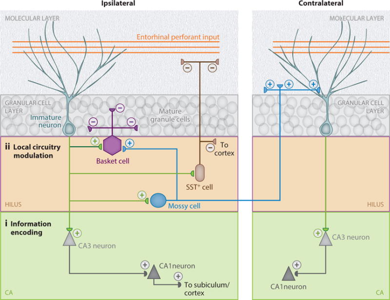
Circuitry properties of newborn neurons in the adult hippocampus. A schematic illustration of connectivity of newborn neurons in the adult dentate gyrus, highlighting the two modes by which newborn neurons can contribute to hippocampal functions, direct information processing via CA3 neurons (i, direct pathway) and modulation of local circuitry via hilar interneurons and mossy cells (ii, indirect pathway). GCL, granule cell layer; SST+ neurons, somatostatin-expressing interneurons.
In the second mode, newborn neurons could actively modulate local circuit activity, for example, maintaining a basal tone of excitation/inhibition or facilitating information encoding by increasing signal to noise in the region and/or by priming circuits to respond (Figure 2). Immature newborn neurons target hilar basket cells (Toni et al. 2008), which provide strong inhibition of a large number of mature granule cells and regulate network oscillations (Freund 2003). Indeed, in vivo recordings from the dentate gyrus in anesthetized mice have shown that elimination of adult neurogenesis leads to decreased amplitude of perforant path–evoked responses and a marked increase in both the amplitude of spontaneous γ-frequency bursts in the dentate gyrus and the synchronization of dentate neuron firing to these bursts (Lacefield et al. 2012). Immediate early gene expression analysis has also shown increased activation of dentate granule cells in response to specific learning tasks after ablation of adult neurogenesis (Burghardt et al. 2012). In addition to interneurons, newborn neurons also innervate hilar mossy cells (Toni et al. 2008), which are glutamatergic and activate local interneurons and contralateral newborn and mature granule cells. Together, a modulator mode provides an amplification mechanism that allows a small number of newborn neurons to impact the global function of the dentate gyrus across both hemispheres. In addition, hippocampal somatostatin-expressing interneurons, including those in the hilus, send distal projections and directly modulate inhibition in the entorhinal cortex (Melzer et al. 2012). This finding raises the intriguing possibility that newborn neurons shape network properties well beyond the dentate gyrus by regulating these long projection interneurons. More studies are needed to investigate potential roles of new neurons as an active modulator of proximal and distal circuitry, especially in awake behaving animals.
Adult neurogenesis could also contribute indirectly to brain functions through alternations in structure properties of the circuitry, a possibility that has been rarely tested experimentally (Figure 2). For example, adult neurogenesis disrupts existing synapses of mature neurons owing to competition during new-neuron synaptic integration (Toni et al. 2007, 2008). The impact of such structural plasticity of mature neurons on brain functions is not well understood. In addition, adult hippocampal neurogenesis generates astrocytes that migrate into the hilus, the granule cell layer, and the molecular layer (Bonaguidi et al. 2011, Encinas et al. 2011). Given the critical role of astrocytes in regulating various brain functions (Clarke & Barres 2013), including adult hippocampal neurogenesis (Song et al. 2002), this is a largely untapped area that warrants future exploration.
FUNCTIONS OF ADULT HIPPOCAMPAL NEUROGENESIS IN COGNITION AND MOOD REGULATION
Immediately after the initial discovery of neurogenesis in the postnatal rat hippocampus, Altman (1967) postulated that newborn neurons are critical for learning and memory. Studies have since shown that hippocampus-dependent learning and memory (Gould et al. 1999), experience, mood, behavioral states, and antidepressants dynamically regulate multiple adult hippocampal neurogenesis processes (Deng et al. 2010, Sahay & Hen 2007). The first experimental evidence in mammals of a casual role of adult neurogenesis in generation or modification of specific behaviors came from a study in which blockade of neurogenesis in the adult mouse by an antimitotic agent disrupts trace eye-blink conditioning and trace fear conditioning, but not contextual feature conditioning and spatial memory (Shors et al. 2001). Since then, the field has gradually transitioned from correlative studies with manipulations that lack specificity to more sophisticated genetic and optogenetic approaches with enhanced temporal and spatial resolution and targeted behavioral protocols. Computational modeling has also been instrumental in framing possible functions of adult hippocampal neurogenesis and its underlying mechanisms (Aimone & Gage 2011). Although the idea is still under debate, the dorsal and ventral hippocampus are likely involved in fine-tuned, spatially discrete memory processes and affective behaviors, respectively, and adult hippocampal neurogenesis has been implicated in both functions (Deng et al. 2010, Kitabatake et al. 2007, Sahay et al. 2011b).
Cognitive Functions
A central dogma regarding information processing through the trisynaptic circuit of the hippocampus has been that the dentate gyrus mediates pattern separation, the ability to distinguish similar stimuli and contexts, whereas CA3 mediates pattern completion, the reinstatement of activity patterns correlated with complete contexts and associations using only partial or degraded information. Building on Marr’s description of the information-processing capacity of the hippocampus based on its structure and intrinsic connectivity, an emergent model proposed that the densely packed dentate gyrus can support the orthogonalization of inputs arriving from the entorhinal cortex (pattern separation) (Marr 1971, Treves & Rolls 1994). The dentate gyrus, in turn, directly projects to CA3, a hippocampal subregion with extensive recurrent connections and thus the putative capacity to reactivate stored patterns using partial inputs (pattern completion). The idea that the dentate gyrus could support pattern separation received empirical support following the observation that cells in the dentate gyrus region are sparsely activated and have very low firing rates. Many behavioral assays of the dentate gyrus and CA3 function have been designed to test this hypothesis, and indeed results suggest that each of these regions may play a role in pattern separation and completion, respectively (Hunsaker & Kesner 2013). Recent evidence also supports an essential role for newborn granule neurons in mediating pattern separation, as inferred from behavioral deficits in mnemonic discrimination when adult neurogenesis is impaired (Aimone et al. 2011, Sahay et al. 2011b). For example, ablation of adult neurogenesis via irradiation impairs the ability of the mice to discriminate stimuli with little spatial separation, but not stimuli widely separated in space, in two spatial memory tasks (Clelland et al. 2009). Conversely, increasing the number of adult-born neurons by deleting the proapoptotic gene Bax from adult neural precursors and their progeny enhances their ability to differentiate between overlapping contextual representations (Sahay et al. 2011a). In one striking example, mice engineered to block synaptic release via tetanus toxin light-chain expression in mature dentate granule cells, but not in most immature neurons younger than four weeks, exhibit improved discrimination of very similar contexts in a fear-conditioning test, and blockade of adult neurogenesis via irradiation impairs context discrimination, suggesting a predominant role of immature neurons in mediating pattern separation (Nakashiba et al. 2012). However, these mice exhibit defects in the water-maze task and fear-conditioning tasks with partial cues presented, which suggests a critical role for mature granule cells in rapid pattern completion. One must recognize that the computational definition of pattern separation and completion implies a strict input–output relationship at the neural level that is not necessarily congruent with the use of this concept to describe behavior (Santoro 2013). It is still not clear, for example, that an animal that appears to pattern complete by behaving indistinguishably in a full-cue versus partial-cue context is doing so by using attractor dynamics in the CA3 region or by engaging mechanisms that compensate for the activation of only a subset of synapses to recruit the full complement of synapses involved in encoding (Knierim & Zhang 2012). Likewise, it is not clear that similar contexts are encoded by minimally overlapping ensembles of dentate granule cells.
Adult hippocampal neurogenesis has also been implicated in other aspects of contextual and spatial memory (Deng et al. 2010). By varying the timing between adult neurogenesis ablation and behavioral tests, studies have also pinpointed essential roles of adult-born immature neurons at various stages for these functions, especially when the task is difficult (Deng et al. 2009, Denny et al. 2012). These results corroborate findings of special cellular properties associated with immature stages and support the immature neuron model of the contribution of adult neurogenesis to brain functions. Adult neurogenesis has also been implicated in memory consolidation and the reorganization of memory traces to extrahippocampal structures, such as the prefrontal cortex (Kitamura et al. 2009). One study has shown that decreased adult hippocampal neurogenesis is accompanied by a prolonged period of hippocampus-dependent associative fear memory, whereas increased adult neurogenesis is associated with accelerated reorganization of memory traces that rely less on the hippocampus. In another interesting study, longitudinal activity data were collected on a large number of inbred mice, which shared one large enriched environment, to explore the relationship among cognitive challenges, adult hippocampal neurogenesis, and the development of individual behavioral traits. The size of the roaming area explored by an individual mouse was positively correlated with the amount of hippocampal neurogenesis (Freund et al. 2013), suggesting that one function of adult neurogenesis may be to shape the neuronal circuitry according to individual needs and improve adaptability over the life course of the individual.
There are many inconsistencies in the current literature regarding effects of various manipulations of adult neurogenesis levels on behavioral test outcomes, which have been summarized in previous reviews (Deng et al. 2010, W.R. Kim et al. 2012). Many possible factors contribute to these contradictory findings, such as differences in the genetic background, experimental manipulation, and behavioral paradigms. One major limitation of traditional approaches is the chronic nature of manipulations that affect adult neurogenesis throughout multiple phases of the learning process; therefore, it is not always clear whether adult-born neurons contribute to encoding, consolidation, storage, and/or retrieval processes. In addition, compensatory network changes may occur following ablation of adult neurogenesis (Singer et al. 2011). Newly available tools now allow for specific manipulation of the activity of adult-born neurons at distinct stages of maturation and during specific stages of learning and recall (W.R. Kim et al. 2012). One recent study of retrovirally targeted newborn neurons in the adult mouse dentate gyrus showed that optogenetic suppression of four-week-old, but not two- or eight-week-old, newborn neurons during recall trials impairs contextual fear memory and spatial memory retrieval (Gu et al. 2012). Given the increasing availability of sophisticated genetic models to target specific populations of neural progenitor subtypes or newborn neurons at specific maturation stages, combined with optogenetic and pharmacogenetic tools to manipulate neuronal activity with spatial and temporal precision, future studies will be able to directly address questions of how and when newborn neurons contribute to neural function.
Mood Regulation
It is now well-established that stress negatively regulates progenitor proliferation and new-neuron survival (Gould et al. 1992), whereas clinical antidepressant treatments, including electroconvulsive therapy and chemical antidepressants (Malberg et al. 2000), promote proliferation of neural progenitors and maturation of newborn neurons during adult hippocampal neurogenesis (Sahay & Hen 2007, Warner-Schmidt & Duman 2006). Such effects are evolutionarily conserved from rodents to nonhuman primates (Perera et al. 2007) and potentially to humans (Boldrini et al. 2009). Ablation of adult neurogenesis does not appear to alter affective phenotypes at basal levels but abolishes some antidepressant-induced behaviors in rodents (Santarelli et al. 2003) and nonhuman primates (Perera et al. 2011). Emerging evidence suggests a critical role of adult hippocampal neurogenesis in the stress response by suppressing the hypothalamic-pituitary-adrenal (HPA) axis. In mice with adult neurogenesis ablated, mild stress leads to increased levels of stress hormones and greater stress responses in behavioral tests (Schloesser et al. 2009, Snyder et al. 2011). Furthermore, blockade of adult neurogenesis abolishes the antidepressant effect of hippocampal regulation of the HPA axis after chronic stress (Surget et al. 2011). The mechanism by which adult-born neurons regulate the HPA axis under basal conditions and upon antidepressant treatment remains to be determined. Anatomical studies have revealed that whereas the dorsal hippocampus projects primarily to cortical areas that mediate cognitive processes such as learning and memory and navigation and exploration, the ventral hippocampus projects to the limbic system, including the amygdala, the nucleus accumbens, and the hypothalamus (Fanselow & Dong 2010). Future studies are needed to address how adult-born neurons, especially those in ventral regions, influence neural pathways involved in emotional experience and affective states. It will also be interesting to test the immature neuron model of adult neurogenesis function in the context of mood regulation. Development of optogenetic and pharmacogenetic approaches to target newborn neurons at specific maturation stages in the dorsal or ventral dentate gyrus will facilitate these efforts (Kheirbek et al. 2013).
DYSFUNCTION OF ADULT HIPPOCAMPAL NEUROGENESIS IN BRAIN DISORDERS
The dentate gyrus is vulnerable to cell death; but as one of the most labile structures in the brain in terms of population dynamics, it is also subject to the consequences of dysregulated adult neurogenesis. A substantial body of literature addresses changes of adult hippocampal neurogenesis in rodents, and limited reports in humans, in the context of various pathophysiological conditions, including aging, epilepsy, stroke, degenerative neurological disorders, and neuropsychiatric disorders (Kempermann et al. 2008, Parent 2003, Sahay & Hen 2007, Winner et al. 2011). In most cases, whether these changes represent adaptive responses to various pathophysiological conditions, or are part of the pathophysiology that contributes to the condition, is unknown. Examples in animal models now suggest that dysfunction of adult hippocampal neurogenesis may play a causal role in brain disorders. There are two modes by which dysregulated adult hippocampal neurogenesis can contribute to dysfunction of the hippocampus: a loss-of-function mode due to decreased new-neuron production and integration, and a gain-of-function mode due to aberrant development and integration of new neurons.
Dysfunction via Loss-of-Function
Fragile X syndrome, the most common form of inherited intellectual disability, is caused by the functional loss of fragile X mental retardation protein (FMRP). Fmrp null mice exhibit deficits in some forms of hippocampus-dependent learning, accompanied by reduced adult hippocampal neurogenesis due to impaired neuronal differentiation and survival (Luo et al. 2010). Deletion of Fmrp specifically in adult neural progenitor cells using nestin-CreERT2 mice recapitulates defects in both adult neurogenesis and hippocampus-dependent learning, and furthermore, restoration of Fmrp expression in adult neural progenitors alone is sufficient to rescue learning deficits in Fmrp null mice (Guo et al. 2011). These striking results suggest a causal role of adult neurogenesis dysfunction in learning impairments associated with fragile X syndrome, at least in animal models. Whether this is generalizable to other disorders remains to be seen.
One major regulator of adult hippocampal neurogenesis is aging. Over the subject’s lifetime, the rate of adult neurogenesis decreases dramatically, from rodents to primates, which may contribute to the dysfunction of hippocampus (Ming & Song 2011). The rate of decline of adult neurogenesis during aging is much more robust in rodents (about tenfold) than in humans (about fourfold) (Spalding et al. 2013).
Dysfunction via Gain-of-Function
Dentate granule cells may play a central role in the pathogenesis of temporal lobe epilepsy, one of the most common human seizure-related disorders (Houser 1992). In animal models of epilepsy, pilocarpine-induced status epilepticus leads to a dramatic and prolonged increase in dentate neural progenitor proliferation (Parent et al. 1997). However, many of these newborn neurons integrate aberrantly, displaying hilar basal dendrites with spines, ectopic hilar localization of the cell body, and mossy fiber sprouting (Jessberger et al. 2007, Kron et al. 2010), similar to what has been observed in postmortem dentate gyri of patients with temporal lobe epilepsy (Houser 1992). Eliminating cohorts of newborn neurons decreases status epilepticus–induced mossy fiber sprouting and ectopic granule cells (Kron et al. 2010) and attenuates spontaneous recurrent seizures in mice (Jung et al. 2004). Separately, deletion of PTEN (phosphatase and tensin homolog deleted on chromosome ten) in a small percentage of dentate granule cells born postnatally is sufficient to cause spontaneous seizure within four weeks, accompanied with aberrant granule cell morphology seen in epilepsy (Pun et al. 2012). Collectively, these studies provide strong evidence that dysfunction of adult hippocampal neurogenesis plays a causal role in epileptogenesis.
In another gain-of-function example, retrovirus-mediated knockdown of Disrupted in Schizophrenia I (DISC1), a risk gene for major mental illness (Thomson et al. 2013), leads to aberrant integration of newborn dentate granule neurons in the adult mouse hippocampus, including ectopic location of the cell body to the outer granule cell layer and molecular layer, aberrant axonal targeting beyond CA3, hyperexcitability, and aberrant formation of synaptic inputs and outputs, due in part to hyperactivation of the mTOR pathway in newborn neurons (Duan et al. 2007; Faulkner et al. 2008; J.Y. Kim et al. 2009, 2012). Dysregulated adult hippocampal neurogenesis following DISC1 knockdown in one cohort of newborn neurons is sufficient to cause several behavioral phenotypes, including pronounced learning and memory deficits (in the object-place recognition task and the spatial version of the Morris water maze), as well as clear anxiety and depression-like phenotypes (in the forced-swim test and elevated plus maze) (Zhou et al. 2013). Inactivation of these aberrant neurons reverses specific behavioral phenotypes, indicating a causal role of adult neurogenesis dysfunction in behavioral impairments.
Notably, the impact of adult neurogenesis dysfunction due to gain-of-function on animal behavior is generally more pronounced than that seen in loss-of-function conditions. This finding may not be surprising because the complete absence or removal of a system may trigger the recruitment of alternative pathways to compensate (Singer et al. 2011), whereas miswiring of newborn neurons can be more detrimental, especially given their high excitability and unique properties. These findings have significant implications for future cell-replacement therapy in which correct wiring of transplanted neurons could be essential for functional benefits and for avoiding potential side effects.
CONCLUSION
In the past decade we have witnessed rapid advances in the adult neurogenesis field, with significant progress in (a) the characterization of this phenomenon in different species, including humans; (b) the delineation of neurogenic processes and properties of adult-born neurons; (c) exploration of its function at circuitry and behavioral levels; and (d) an appreciation of how dysfunction of adult neurogenesis may contribute to brain disorders. Despite these tremendous findings, understanding the function of adult hippocampal neurogenesis remains a central goal in the field. Perhaps one of most frequently asked questions is why it occurs in the dentate gyrus. The dentate gyrus is one of the two regions with continuous neurogenesis from rodents to humans. Fully addressing these questions will require a multidisciplinary approach and new technologies. First, we need to know more about basic properties of the dentate gyrus and how it processes information and contributes to hippocampal functions, which will provide the framework to delineate the contribution of adult neurogenesis. Second, we need to have a better knowledge of the dentate circuitry, especially synaptic outputs of newborn neurons. Third, recordings of newborn neurons at different maturation stages in awake behaving animals will provide critical information to test current models of how adult-born neurons contribute to brain functions and dysfunctions. Fourth, the field needs to address contradictory results from behavioral analyses using newly available tools with better cell-type specificity and higher temporal and spatial resolution. Fifth, we need to consider how other plasticity associated with adult neurogenesis also contributes to brain functions, such as the generation of new astrocytes and induced structural changes in mature neurons. Sixth, comparative studies of two primary neurogenic regions in different species have proven highly informative, and ultimately, we want to understand the function of adult neurogenesis in humans.
It was originally proposed that adult neurogenesis is not a cell-replacement mechanism in which dying individual neurons are functionally replaced by new neurons, but instead continuously provides new cohorts of immature neurons with properties and information-processing capacities that are distinct from those of existing mature neurons (Ge et al. 2007). This immature neuron model of the adult neurogenesis contribution to brain function has gained significant support over the past few years from additional comparisons of immature and mature neurons, computational modeling, and animal behavioral analyses. Building on this model is the plastic dentate gyrus hypothesis: Adult neurogenesis represents a continuous developmental process that maintains a highly plastic dentate circuitry, collectively with the addition of immature neurons with unique properties and new astrocytes and with the continued structural plasticity of associated mature neurons in broader brain regions. The heterogeneous nature of the dentate gyrus, with a small immature neuronal cohort that is highly plastic and excitable and with a large population of mature granule cells that are sparsely activated with high input specificity, offers unique information-processing power that can adapt to dynamic needs over the lifetime.
Understanding the physiological function of adult neurogenesis not only provides a new prospective on the plasticity of the mature nervous system, but also has significant implications for our understanding of several brain disorders and regenerative medicine. Recent evidence supports a critical contribution of dysfunctional postnatal neurogenesis, via both loss-of-function and gain-of-function modes, to developmental disorders and may be a crucial mechanism that initiates the onset of disorders such as autism and schizophrenia (Ming & Song 2009). Recent animal model studies have suggested that treating molecular deficits underlying neurodevelopmental disorders could result in significant amelioration of associated behavioral phenotypes, even when treatments were initiated in adults (Ehninger et al. 2008). Therefore, targeting adult neurogenesis could be a novel potential therapeutic strategy for these disorders. Basic principles learned from normal and dysregulated adult neuronal development and synaptic integration of newborn neurons will also provide invaluable information for the future development of cell-replacement therapy.
Acknowledgments
We apologize that we could not cite all primary literatures owing to space limitations. The research in the authors’ laboratories was supported by National Institutes of Health (R01NS048271, R01HD069184 to G-l.M. & R37NS047344, R21ES021957 to H.S.), the Maryland Stem Cell Research Fund (K.C. & G-l.M.), NARSAD (K.C. & G-l.M.), the Simons Foundation (H.S.), and the Dr. Miriam and Sheldon G. Adelson Medical Research Foundation (G-l.M.).
Footnotes
DISCLOSURE STATEMENT
The authors are not aware of any affiliations, memberships, funding, or financial holdings that might be perceived as affecting the objectivity of this review.
LITERATURE CITED
- Aimone JB, Deng W, Gage FH. Resolving new memories: a critical look at the dentate gyrus, adult neurogenesis, and pattern separation. Neuron. 2011;70:589–96. doi: 10.1016/j.neuron.2011.05.010. [DOI] [PMC free article] [PubMed] [Google Scholar]
- Aimone JB, Gage FH. Modeling new neuron function: a history of using computational neuroscience to study adult neurogenesis. Eur J Neurosci. 2011;33:1160–69. doi: 10.1111/j.1460-9568.2011.07615.x. [DOI] [PubMed] [Google Scholar]
- Altman J. In: The Neurosciences: Second Study Program. Quarton GC, Melnechuck T, Schmitt FO, editors. New York: Rockefeller Univ. Press; 1967. pp. 723–43. [Google Scholar]
- Boldrini M, Underwood MD, Hen R, Rosoklija GB, Dwork AJ, et al. Antidepressants increase neural progenitor cells in the human hippocampus. Neuropsychopharmacology. 2009;34:2376–89. doi: 10.1038/npp.2009.75. [DOI] [PMC free article] [PubMed] [Google Scholar]
- Bonaguidi MA, Song J, Ming GL, Song H. A unifying hypothesis on mammalian neural stem cell properties in the adult hippocampus. Curr Opin Neurobiol. 2012;22:754–61. doi: 10.1016/j.conb.2012.03.013. [DOI] [PMC free article] [PubMed] [Google Scholar]
- Bonaguidi MA, Wheeler MA, Shapiro JS, Stadel RP, Sun GJ, et al. In vivo clonal analysis reveals self-renewing and multipotent adult neural stem cell characteristics. Cell. 2011;145:1142–55. doi: 10.1016/j.cell.2011.05.024. [DOI] [PMC free article] [PubMed] [Google Scholar]
- Braun SMG, Jessberger S. Adult neurogenesis in the mammalian brain. Front Biol. 2013;8:295–304. [Google Scholar]
- Brus M, Keller M, Lévy F. Temporal features of adult neurogenesis: differences and similarities across mammalian species. Front Neurosci. 2013;7:135. doi: 10.3389/fnins.2013.00135. [DOI] [PMC free article] [PubMed] [Google Scholar]
- Burghardt NS, Park EH, Hen R, Fenton AA. Adult-born hippocampal neurons promote cognitive flexibility in mice. Hippocampus. 2012;22:1795–808. doi: 10.1002/hipo.22013. [DOI] [PMC free article] [PubMed] [Google Scholar]
- Cabezas C, Irinopoulou T, Cauli B, Poncer JC. Molecular and functional characterization of GAD67-expressing, newborn granule cells in mouse dentate gyrus. Front Neural Circuits. 2013;7:60. doi: 10.3389/fncir.2013.00060. [DOI] [PMC free article] [PubMed] [Google Scholar]
- Cabezas C, Irinopoulou T, Gauvain G, Poncer JC. Presynaptic but not postsynaptic GABA signaling at unitary mossy fiber synapses. J Neurosci. 2012;32:11835–40. doi: 10.1523/JNEUROSCI.5543-11.2012. [DOI] [PMC free article] [PubMed] [Google Scholar]
- Cameron HA, McKay RD. Adult neurogenesis produces a large pool of new granule cells in the dentate gyrus. J Comp Neurol. 2001;435:406–17. doi: 10.1002/cne.1040. [DOI] [PubMed] [Google Scholar]
- Chancey JH, Adlaf EW, Sapp MC, Pugh PC, Wadiche JI, Overstreet-Wadiche LS. GABA depolarization is required for experience-dependent synapse unsilencing in adult-born neurons. J Neurosci. 2013;33:6614–22. doi: 10.1523/JNEUROSCI.0781-13.2013. [DOI] [PMC free article] [PubMed] [Google Scholar]
- Chancey JH, Poulsen DJ, Wadiche JI, Overstreet-Wadiche L. Hilar mossy cells provide the first glutamatergic synapses to adult-born dentate granule cells. J Neurosci. 2014;34:2349–54. doi: 10.1523/JNEUROSCI.3620-13.2014. [DOI] [PMC free article] [PubMed] [Google Scholar]
- Clarke LE, Barres BA. Emerging roles of astrocytes in neural circuit development. Nat Rev Neurosci. 2013;14:311–21. doi: 10.1038/nrn3484. [DOI] [PMC free article] [PubMed] [Google Scholar]
- Clelland CD, Choi M, Romberg C, Clemenson GD, Jr, Fragniere A, et al. A functional role for adult hippocampal neurogenesis in spatial pattern separation. Science. 2009;325:210–13. doi: 10.1126/science.1173215. [DOI] [PMC free article] [PubMed] [Google Scholar]
- Deng W, Aimone JB, Gage FH. New neurons and new memories: How does adult hippocampal neurogenesis affect learning and memory? Nat Rev Neurosci. 2010;11:339–50. doi: 10.1038/nrn2822. [DOI] [PMC free article] [PubMed] [Google Scholar]
- Deng W, Saxe MD, Gallina IS, Gage FH. Adult-born hippocampal dentate granule cells undergoing maturation modulate learning and memory in the brain. J Neurosci. 2009;29:13532–42. doi: 10.1523/JNEUROSCI.3362-09.2009. [DOI] [PMC free article] [PubMed] [Google Scholar]
- Denny CA, Burghardt NS, Schachter DM, Hen R, Drew MR. 4- to 6-week-old adult-born hippocampal neurons influence novelty-evoked exploration and contextual fear conditioning. Hippocampus. 2012;22:1188–201. doi: 10.1002/hipo.20964. [DOI] [PMC free article] [PubMed] [Google Scholar]
- Deshpande A, Bergami M, Ghanem A, Conzelmann KK, Lepier A, et al. Retrograde monosynaptic tracing reveals the temporal evolution of inputs onto new neurons in the adult dentate gyrus and olfactory bulb. Proc Natl Acad Sci USA. 2013;110:E1152–61. doi: 10.1073/pnas.1218991110. [DOI] [PMC free article] [PubMed] [Google Scholar]
- Dhaliwal J, Lagace DC. Visualization and genetic manipulation of adult neurogenesis using transgenic mice. Eur J Neurosci. 2011;33:1025–36. doi: 10.1111/j.1460-9568.2011.07600.x. [DOI] [PubMed] [Google Scholar]
- Dieni CV, Nietz AK, Panichi R, Wadiche JI, Overstreet-Wadiche L. Distinct determinants of sparse activation during granule cell maturation. J Neurosci. 2013;33:19131–42. doi: 10.1523/JNEUROSCI.2289-13.2013. [DOI] [PMC free article] [PubMed] [Google Scholar]
- Duan X, Chang JH, Ge S, Faulkner RL, Kim JY, et al. Disrupted-In-Schizophrenia 1 regulates integration of newly generated neurons in the adult brain. Cell. 2007;130:1146–58. doi: 10.1016/j.cell.2007.07.010. [DOI] [PMC free article] [PubMed] [Google Scholar]
- Ehninger D, Li W, Fox K, Stryker MP, Silva AJ. Reversing neurodevelopmental disorders in adults. Neuron. 2008;60:950–60. doi: 10.1016/j.neuron.2008.12.007. [DOI] [PMC free article] [PubMed] [Google Scholar]
- Encinas JM, Michurina TV, Peunova N, Park J-H, Tordo J, et al. Division-coupled astrocytic differentiation and age-related depletion of neural stem cells in the adult hippocampus. Cell Stem Cell. 2011;8:566–79. doi: 10.1016/j.stem.2011.03.010. [DOI] [PMC free article] [PubMed] [Google Scholar]
- Ernst A, Alkass K, Bernard S, Salehpour M, Perl S, et al. Neurogenesis in the striatum of the adult human brain. Cell. 2014;156:1072–83. doi: 10.1016/j.cell.2014.01.044. [DOI] [PubMed] [Google Scholar]
- Espósito MS, Piatti VC, Laplagne DA, Morgenstern NA, Ferrari CC, et al. Neuronal differentiation in the adult hippocampus recapitulates embryonic development. J Neurosci. 2005;25:10074–86. doi: 10.1523/JNEUROSCI.3114-05.2005. [DOI] [PMC free article] [PubMed] [Google Scholar]
- Fanselow MS, Dong HW. Are the dorsal and ventral hippocampus functionally distinct structures? Neuron. 2010;65:7–19. doi: 10.1016/j.neuron.2009.11.031. [DOI] [PMC free article] [PubMed] [Google Scholar]
- Faulkner RL, Jang M-H, Liu X-B, Duan X, Sailor KA, et al. Development of hippocampal mossy fiber synaptic outputs by new neurons in the adult brain. Proc Natl Acad Sci USA. 2008;105:14157–62. doi: 10.1073/pnas.0806658105. [DOI] [PMC free article] [PubMed] [Google Scholar]
- Freund J, Brandmaier AM, Lewejohann L, Kirste I, Kritzler M, et al. Emergence of individuality in genetically identical mice. Science. 2013;340:756–59. doi: 10.1126/science.1235294. [DOI] [PubMed] [Google Scholar]
- Freund TF. Interneuron Diversity series: Rhythm and mood in perisomatic inhibition. Trends Neurosci. 2003;26:489–95. doi: 10.1016/S0166-2236(03)00227-3. [DOI] [PubMed] [Google Scholar]
- Gage FH, Temple S. Neural stem cells: generating and regenerating the brain. Neuron. 2013;80:588–601. doi: 10.1016/j.neuron.2013.10.037. [DOI] [PubMed] [Google Scholar]
- Gasbarri A, Sulli A, Packard MG. The dopaminergic mesencephalic projections to the hippocampal formation in the rat. Prog Neuropsychopharmacol Biol Psychiatry. 1997;21:1–22. doi: 10.1016/s0278-5846(96)00157-1. [DOI] [PubMed] [Google Scholar]
- Ge S, Goh EL, Sailor KA, Kitabatake Y, Ming GL, Song H. GABA regulates synaptic integration of newly generated neurons in the adult brain. Nature. 2006;439:589–93. doi: 10.1038/nature04404. [DOI] [PMC free article] [PubMed] [Google Scholar]
- Ge S, Sailor KA, Ming GL, Song H. Synaptic integration and plasticity of new neurons in the adult hippocampus. J Physiol. 2008;586:3759–65. doi: 10.1113/jphysiol.2008.155655. [DOI] [PMC free article] [PubMed] [Google Scholar]
- Ge S, Yang CH, Hsu KS, Ming GL, Song H. A critical period for enhanced synaptic plasticity in newly generated neurons of the adult brain. Neuron. 2007;54:559–66. doi: 10.1016/j.neuron.2007.05.002. [DOI] [PMC free article] [PubMed] [Google Scholar]
- Göritz C, Frisén J. Neural stem cells and neurogenesis in the adult. Cell Stem Cell. 2012;10:657–59. doi: 10.1016/j.stem.2012.04.005. [DOI] [PubMed] [Google Scholar]
- Gould E, Beylin A, Tanapat P, Reeves A, Shors TJ. Learning enhances adult neurogenesis in the hippocampal formation. Nat Neurosci. 1999;2:260–65. doi: 10.1038/6365. [DOI] [PubMed] [Google Scholar]
- Gould E, Cameron HA, Daniels DC, Woolley CS, McEwen BS. Adrenal hormones suppress cell division in the adult rat dentate gyrus. J Neurosci. 1992;12:3642–50. doi: 10.1523/JNEUROSCI.12-09-03642.1992. [DOI] [PMC free article] [PubMed] [Google Scholar]
- Gu Y, Arruda-Carvalho M, Wang J, Janoschka SR, Josselyn SA, et al. Optical controlling reveals time-dependent roles for adult-born dentate granule cells. Nat Neurosci. 2012;15:1700–6. doi: 10.1038/nn.3260. [DOI] [PMC free article] [PubMed] [Google Scholar]
- Guo W, Allan AM, Zong R, Zhang L, Johnson EB, et al. Ablation of Fmrp in adult neural stem cells disrupts hippocampus-dependent learning. Nat Med. 2011;17:559–65. doi: 10.1038/nm.2336. [DOI] [PMC free article] [PubMed] [Google Scholar]
- Houser CR. Morphological changes in the dentate gyrus in human temporal lobe epilepsy. Epilepsy Res Suppl. 1992;7:223–34. [PubMed] [Google Scholar]
- Hunsaker MR, Kesner RP. The operation of pattern separation and pattern completion processes associated with different attributes or domains of memory. Neurosci Biobehav Rev. 2013;37:36–58. doi: 10.1016/j.neubiorev.2012.09.014. [DOI] [PubMed] [Google Scholar]
- Jessberger S, Zhao C, Toni N, Clemenson GD, Jr, Li Y, Gage FH. Seizure-associated, aberrant neurogenesis in adult rats characterized with retrovirus-mediated cell labeling. J Neurosci. 2007;27:9400–7. doi: 10.1523/JNEUROSCI.2002-07.2007. [DOI] [PMC free article] [PubMed] [Google Scholar]
- Jung KH, Chu K, Kim M, Jeong SW, Song YM, et al. Continuous cytosine-b-D-arabinofuranoside infusion reduces ectopic granule cells in adult rat hippocampus with attenuation of spontaneous recurrent seizures following pilocarpine-induced status epilepticus. Eur J Neurosci. 2004;19:3219–26. doi: 10.1111/j.0953-816X.2004.03412.x. [DOI] [PubMed] [Google Scholar]
- Kempermann G, Gage FH. New nerve cells for the adult brain. Sci Am. 1999;280:48–53. doi: 10.1038/scientificamerican0599-48. [DOI] [PubMed] [Google Scholar]
- Kempermann G, Krebs J, Fabel K. The contribution of failing adult hippocampal neurogenesis to psychiatric disorders. Curr Opin Psychiatry. 2008;21:290–95. doi: 10.1097/YCO.0b013e3282fad375. [DOI] [PubMed] [Google Scholar]
- Kheirbek MA, Drew LJ, Burghardt NS, Costantini DO, Tannenholz L, et al. Differential control of learning and anxiety along the dorsoventral axis of the dentate gyrus. Neuron. 2013;77:955–68. doi: 10.1016/j.neuron.2012.12.038. [DOI] [PMC free article] [PubMed] [Google Scholar]
- Kim JY, Duan X, Liu CY, Jang M-H, Guo JU, et al. DISC1 regulates new neuron development in the adult brain via modulation of AKT-mTOR signaling through KIAA1212. Neuron. 2009;63:761–73. doi: 10.1016/j.neuron.2009.08.008. [DOI] [PMC free article] [PubMed] [Google Scholar]
- Kim JY, Liu CY, Zhang F, Duan X, Wen Z, et al. Interplay between DISC1 and GABA signaling regulates neurogenesis in mice and risk for schizophrenia. Cell. 2012;148:1051–64. doi: 10.1016/j.cell.2011.12.037. [DOI] [PMC free article] [PubMed] [Google Scholar]
- Kim WR, Christian K, Ming GL, Song H. Time-dependent involvement of adult-born dentate granule cells in behavior. Behav Brain Res. 2012;227:470–79. doi: 10.1016/j.bbr.2011.07.012. [DOI] [PMC free article] [PubMed] [Google Scholar]
- Kitabatake Y, Sailor KA, Ming GL, Song H. Adult neurogenesis and hippocampal memory function: new cells, more plasticity, new memories? Neurosurg Clin N Am. 2007;18:105–13. x. doi: 10.1016/j.nec.2006.10.008. [DOI] [PMC free article] [PubMed] [Google Scholar]
- Kitamura T, Saitoh Y, Takashima N, Murayama A, Niibori Y, et al. Adult neurogenesis modulates the hippocampus-dependent period of associative fear memory. Cell. 2009;139:814–27. doi: 10.1016/j.cell.2009.10.020. [DOI] [PubMed] [Google Scholar]
- Knierim JJ, Zhang K. Attractor dynamics of spatially correlated neural activity in the limbic system. Annu Rev Neurosci. 2012;35:267–85. doi: 10.1146/annurev-neuro-062111-150351. [DOI] [PMC free article] [PubMed] [Google Scholar]
- Kohler SJ, Williams NI, Stanton GB, Cameron JL, Greenough WT. Maturation time of new granule cells in the dentate gyrus of adult macaque monkeys exceeds six months. Proc Natl Acad Sci USA. 2011;108:10326–31. doi: 10.1073/pnas.1017099108. [DOI] [PMC free article] [PubMed] [Google Scholar]
- Kriegstein A, Alvarez-Buylla A. The glial nature of embryonic and adult neural stem cells. Annu Rev Neurosci. 2009;32:149–84. doi: 10.1146/annurev.neuro.051508.135600. [DOI] [PMC free article] [PubMed] [Google Scholar]
- Kron MM, Zhang H, Parent JM. The developmental stage of dentate granule cells dictates their contribution to seizure-induced plasticity. J Neurosci. 2010;30:2051–59. doi: 10.1523/JNEUROSCI.5655-09.2010. [DOI] [PMC free article] [PubMed] [Google Scholar]
- Kumamoto N, Gu Y, Wang J, Janoschka S, Takemaru K, et al. A role for primary cilia in glutamatergic synaptic integration of adult-born neurons. Nat Neurosci. 2012;15:399–405, S1. doi: 10.1038/nn.3042. [DOI] [PMC free article] [PubMed] [Google Scholar]
- Lacefield CO, Itskov V, Reardon T, Hen R, Gordon JA. Effects of adult-generated granule cells on coordinated network activity in the dentate gyrus. Hippocampus. 2012;22:106–16. doi: 10.1002/hipo.20860. [DOI] [PMC free article] [PubMed] [Google Scholar]
- Laplagne DA, Espósito MS, Piatti VC, Morgenstern NA, Zhao C, et al. Functional convergence of neurons generated in the developing and adult hippocampus. PLoS Biol. 2006;4:e409. doi: 10.1371/journal.pbio.0040409. [DOI] [PMC free article] [PubMed] [Google Scholar]
- Laplagne DA, Kamienkowski JE, Espósito MS, Piatti VC, Zhao C, et al. Similar GABAergic inputs in dentate granule cells born during embryonic and adult neurogenesis. Eur J Neurosci. 2007;25:2973–81. doi: 10.1111/j.1460-9568.2007.05549.x. [DOI] [PubMed] [Google Scholar]
- Lemaire V, Tronel S, Montaron MF, Fabre A, Dugast E, Abrous DN. Long-lasting plasticity of hippocampal adult-born neurons. J Neurosci. 2012;32:3101–8. doi: 10.1523/JNEUROSCI.4731-11.2012. [DOI] [PMC free article] [PubMed] [Google Scholar]
- Lepousez G, Valley MT, Lledo PM. The impact of adult neurogenesis on olfactory bulb circuits and computations. Annu Rev Physiol. 2013;75:339–63. doi: 10.1146/annurev-physiol-030212-183731. [DOI] [PubMed] [Google Scholar]
- Leutgeb JK, Leutgeb S, Moser MB, Moser EI. Pattern separation in the dentate gyrus and CA3 of the hippocampus. Science. 2007;315:961–66. doi: 10.1126/science.1135801. [DOI] [PubMed] [Google Scholar]
- Li Y, Aimone JB, Xu X, Callaway EM, Gage FH. Development of GABAergic inputs controls the contribution of maturing neurons to the adult hippocampal network. Proc Natl Acad Sci USA. 2012;109:4290–95. doi: 10.1073/pnas.1120754109. [DOI] [PMC free article] [PubMed] [Google Scholar]
- Li Y, Stam FJ, Aimone JB, Goulding M, Callaway EM, Gage FH. Molecular layer perforant path-associated cells contribute to feed-forward inhibition in the adult dentate gyrus. Proc Natl Acad Sci USA. 2013;110:9106–11. doi: 10.1073/pnas.1306912110. [DOI] [PMC free article] [PubMed] [Google Scholar]
- Liu YB, Lio PA, Pasternak JF, Trommer BL. Developmental changes in membrane properties and postsynaptic currents of granule cells in rat dentate gyrus. J Neurophysiol. 1996;76:1074–88. doi: 10.1152/jn.1996.76.2.1074. [DOI] [PubMed] [Google Scholar]
- Lledo PM, Alonso M, Grubb MS. Adult neurogenesis and functional plasticity in neuronal circuits. Nat Rev Neurosci. 2006;7:179–93. doi: 10.1038/nrn1867. [DOI] [PubMed] [Google Scholar]
- Luo Y, Shan G, Guo W, Smrt RD, Johnson EB, et al. Fragile X mental retardation protein regulates proliferation and differentiation of adult neural stem/progenitor cells. PLoS Genet. 2010;6:e1000898. doi: 10.1371/journal.pgen.1000898. [DOI] [PMC free article] [PubMed] [Google Scholar]
- Ma DK, Kim WR, Ming GL, Song H. Activity-dependent extrinsic regulation of adult olfactory bulb and hippocampal neurogenesis. Ann N Y Acad Sci. 2009;1170:664–73. doi: 10.1111/j.1749-6632.2009.04373.x. [DOI] [PMC free article] [PubMed] [Google Scholar]
- Malberg JE, Eisch AJ, Nestler EJ, Duman RS. Chronic antidepressant treatment increases neurogenesis in adult rat hippocampus. J Neurosci. 2000;20:9104–10. doi: 10.1523/JNEUROSCI.20-24-09104.2000. [DOI] [PMC free article] [PubMed] [Google Scholar]
- Marín-Burgin A, Mongiat LA, Pardi MB, Schinder AF. Unique processing during a period of high excitation/inhibition balance in adult-born neurons. Science. 2012;335:1238–42. doi: 10.1126/science.1214956. [DOI] [PMC free article] [PubMed] [Google Scholar]
- Markwardt SJ, Dieni CV, Wadiche JI, Overstreet-Wadiche L. Ivy/neurogliaform interneurons coordinate activity in the neurogenic niche. Nat Neurosci. 2011;14:1407–9. doi: 10.1038/nn.2935. [DOI] [PMC free article] [PubMed] [Google Scholar]
- Marr D. Simple memory: a theory for archicortex. Philos Trans R Soc B. 1971;262:23–81. doi: 10.1098/rstb.1971.0078. [DOI] [PubMed] [Google Scholar]
- Melzer S, Michael M, Caputi A, Eliava M, Fuchs EC, et al. Long-range-projecting GABAergic neurons modulate inhibition in hippocampus and entorhinal cortex. Science. 2012;335:1506–10. doi: 10.1126/science.1217139. [DOI] [PubMed] [Google Scholar]
- Ming GL, Song H. Adult neurogenesis in the mammalian central nervous system. Annu Rev Neurosci. 2005;28:223–50. doi: 10.1146/annurev.neuro.28.051804.101459. [DOI] [PubMed] [Google Scholar]
- Ming GL, Song H. DISC1 partners with GSK3β in neurogenesis. Cell. 2009;136:990–92. doi: 10.1016/j.cell.2009.03.005. [DOI] [PMC free article] [PubMed] [Google Scholar]
- Ming GL, Song H. Adult neurogenesis in the mammalian brain: significant answers and significant questions. Neuron. 2011;70:687–702. doi: 10.1016/j.neuron.2011.05.001. [DOI] [PMC free article] [PubMed] [Google Scholar]
- Mongiat LA, Espósito MS, Lombardi G, Schinder AF. Reliable activation of immature neurons in the adult hippocampus. PLoS ONE. 2009;4:e5320. doi: 10.1371/journal.pone.0005320. [DOI] [PMC free article] [PubMed] [Google Scholar]
- Mu Y, Zhao C, Gage FH. Dopaminergic modulation of cortical inputs during maturation of adult-born dentate granule cells. J Neurosci. 2011;31:4113–23. doi: 10.1523/JNEUROSCI.4913-10.2011. [DOI] [PMC free article] [PubMed] [Google Scholar]
- Nakashiba T, Cushman JD, Pelkey KA, Renaudineau S, Buhl DL, et al. Young dentate granule cells mediate pattern separation, whereas old granule cells facilitate pattern completion. Cell. 2012;149:188–201. doi: 10.1016/j.cell.2012.01.046. [DOI] [PMC free article] [PubMed] [Google Scholar]
- Neunuebel JP, Knierim JJ. Spatial firing correlates of physiologically distinct cell types of the rat dentate gyrus. J Neurosci. 2012;32:3848–58. doi: 10.1523/JNEUROSCI.6038-11.2012. [DOI] [PMC free article] [PubMed] [Google Scholar]
- Neunuebel JP, Knierim JJ. CA3 retrieves coherent representations from degraded input: direct evidence for CA3 pattern completion and dentate gyrus pattern separation. Neuron. 2014;81:416–27. doi: 10.1016/j.neuron.2013.11.017. [DOI] [PMC free article] [PubMed] [Google Scholar]
- Overstreet Wadiche L, Bromberg DA, Bensen AL, Westbrook GL. GABAergic signaling to newborn neurons in dentate gyrus. J Neurophysiol. 2005;94:4528–32. doi: 10.1152/jn.00633.2005. [DOI] [PubMed] [Google Scholar]
- Overstreet-Wadiche LS, Bensen AL, Westbrook GL. Delayed development of adult-generated granule cells in dentate gyrus. J Neurosci. 2006a;26:2326–34. doi: 10.1523/JNEUROSCI.4111-05.2006. [DOI] [PMC free article] [PubMed] [Google Scholar]
- Overstreet-Wadiche LS, Bromberg DA, Bensen AL, Westbrook GL. Seizures accelerate functional integration of adult-generated granule cells. J Neurosci. 2006b;26:4095–103. doi: 10.1523/JNEUROSCI.5508-05.2006. [DOI] [PMC free article] [PubMed] [Google Scholar]
- Parent JM. Injury-induced neurogenesis in the adult mammalian brain. Neuroscientist. 2003;9:261–72. doi: 10.1177/1073858403252680. [DOI] [PubMed] [Google Scholar]
- Parent JM, Yu TW, Leibowitz RT, Geschwind DH, Sloviter RS, Lowenstein DH. Dentate granule cell neurogenesis is increased by seizures and contributes to aberrant network reorganization in the adult rat hippocampus. J Neurosci. 1997;17:3727–38. doi: 10.1523/JNEUROSCI.17-10-03727.1997. [DOI] [PMC free article] [PubMed] [Google Scholar]
- Perera TD, Coplan JD, Lisanby SH, Lipira CM, Arif M, et al. Antidepressant-induced neurogenesis in the hippocampus of adult nonhuman primates. J Neurosci. 2007;27:4894–901. doi: 10.1523/JNEUROSCI.0237-07.2007. [DOI] [PMC free article] [PubMed] [Google Scholar]
- Perera TD, Dwork AJ, Keegan KA, Thirumangalakudi L, Lipira CM, et al. Necessity of hippocampal neurogenesis for the therapeutic action of antidepressants in adult nonhuman primates. PLoS ONE. 2011;6:e17600. doi: 10.1371/journal.pone.0017600. [DOI] [PMC free article] [PubMed] [Google Scholar]
- Piatti VC, Davies-Sala MG, Espósito MS, Mongiat LA, Trinchero MF, Schinder AF. The timing for neuronal maturation in the adult hippocampus is modulated by local network activity. J Neurosci. 2011;31:7715–28. doi: 10.1523/JNEUROSCI.1380-11.2011. [DOI] [PMC free article] [PubMed] [Google Scholar]
- Pontes A, Zhang Y, Hu W. Novel functions of GABA signaling in adult neurogenesis. Front Biol. 2013;8:496–507. doi: 10.1007/s11515-013-1270-2. [DOI] [PMC free article] [PubMed] [Google Scholar]
- Pun RY, Rolle IJ, Lasarge CL, Hosford BE, Rosen JM, et al. Excessive activation of mTOR in postnatally generated granule cells is sufficient to cause epilepsy. Neuron. 2012;75:1022–34. doi: 10.1016/j.neuron.2012.08.002. [DOI] [PMC free article] [PubMed] [Google Scholar]
- Ramirez-Amaya V, Marrone DF, Gage FH, Worley PF, Barnes CA. Integration of new neurons into functional neural networks. J Neurosci. 2006;26:12237–41. doi: 10.1523/JNEUROSCI.2195-06.2006. [DOI] [PMC free article] [PubMed] [Google Scholar]
- Ramírez-Amaya V, Vazdarjanova A, Mikhael D, Rosi S, Worley PF, Barnes CA. Spatial exploration-induced Arc mRNA and protein expression: evidence for selective, network-specific reactivation. J Neurosci. 2005;25:1761–68. doi: 10.1523/JNEUROSCI.4342-04.2005. [DOI] [PMC free article] [PubMed] [Google Scholar]
- Sahay A, Hen R. Adult hippocampal neurogenesis in depression. Nat Neurosci. 2007;10:1110–15. doi: 10.1038/nn1969. [DOI] [PubMed] [Google Scholar]
- Sahay A, Scobie KN, Hill AS, O’Carroll CM, Kheirbek MA, et al. Increasing adult hippocampal neurogenesis is sufficient to improve pattern separation. Nature. 2011a;472:466–70. doi: 10.1038/nature09817. [DOI] [PMC free article] [PubMed] [Google Scholar]
- Sahay A, Wilson DA, Hen R. Pattern separation: a common function for new neurons in hippocampus and olfactory bulb. Neuron. 2011b;70:582–88. doi: 10.1016/j.neuron.2011.05.012. [DOI] [PMC free article] [PubMed] [Google Scholar]
- Santarelli L, Saxe M, Gross C, Surget A, Battaglia F, et al. Requirement of hippocampal neurogenesis for the behavioral effects of antidepressants. Science. 2003;301:805–9. doi: 10.1126/science.1083328. [DOI] [PubMed] [Google Scholar]
- Santoro A. Reassessing pattern separation in the dentate gyrus. Front Behav Neurosci. 2013;7:96. doi: 10.3389/fnbeh.2013.00096. [DOI] [PMC free article] [PubMed] [Google Scholar]
- Schloesser RJ, Manji HK, Martinowich K. Suppression of adult neurogenesis leads to an increased hypothalamo-pituitary-adrenal axis response. NeuroReport. 2009;20:553–57. doi: 10.1097/WNR.0b013e3283293e59. [DOI] [PMC free article] [PubMed] [Google Scholar]
- Schmidt-Hieber C, Jonas P, Bischofberger J. Enhanced synaptic plasticity in newly generated granule cells of the adult hippocampus. Nature. 2004;429:184–87. doi: 10.1038/nature02553. [DOI] [PubMed] [Google Scholar]
- Shors TJ, Miesegaes G, Beylin A, Zhao M, Rydel T, Gould E. Neurogenesis in the adult is involved in the formation of trace memories. Nature. 2001;410:372–76. doi: 10.1038/35066584. [DOI] [PubMed] [Google Scholar]
- Sierra A, Encinas JM, Deudero JJ, Chancey JH, Enikolopov G, et al. Microglia shape adult hippocampal neurogenesis through apoptosis-coupled phagocytosis. Cell Stem Cell. 2010;7:483–95. doi: 10.1016/j.stem.2010.08.014. [DOI] [PMC free article] [PubMed] [Google Scholar]
- Singer BH, Gamelli AE, Fuller CL, Temme SJ, Parent JM, Murphy GG. Compensatory network changes in the dentate gyrus restore long-term potentiation following ablation of neurogenesis in young-adult mice. Proc Natl Acad Sci USA. 2011;108:5437–42. doi: 10.1073/pnas.1015425108. [DOI] [PMC free article] [PubMed] [Google Scholar]
- Snyder JS, Kee N, Wojtowicz JM. Effects of adult neurogenesis on synaptic plasticity in the rat dentate gyrus. J Neurophysiol. 2001;85:2423–31. doi: 10.1152/jn.2001.85.6.2423. [DOI] [PubMed] [Google Scholar]
- Snyder JS, Soumier A, Brewer M, Pickel J, Cameron HA. Adult hippocampal neurogenesis buffers stress responses and depressive behaviour. Nature. 2011;476:458–61. doi: 10.1038/nature10287. [DOI] [PMC free article] [PubMed] [Google Scholar]
- Song H, Stevens CF, Gage FH. Astroglia induce neurogenesis from adult neural stem cells. Nature. 2002;417:39–44. doi: 10.1038/417039a. [DOI] [PubMed] [Google Scholar]
- Song J, Sun J, Moss J, Wen Z, Sun GJ, et al. Parvalbumin interneurons mediate neuronal circuitry—neurogenesis coupling in the adult hippocampus. Nat Neurosci. 2013;16:1728–30. doi: 10.1038/nn.3572. [DOI] [PMC free article] [PubMed] [Google Scholar]
- Song J, Zhong C, Bonaguidi MA, Sun GJ, Hsu D, et al. Neuronal circuitry mechanism regulating adult quiescent neural stem-cell fate decision. Nature. 2012;489:150–54. doi: 10.1038/nature11306. [DOI] [PMC free article] [PubMed] [Google Scholar]
- Spalding KL, Bergmann O, Alkass K, Bernard S, Salehpour M, et al. Dynamics of hippocampal neurogenesis in adult humans. Cell. 2013;153:1219–27. doi: 10.1016/j.cell.2013.05.002. [DOI] [PMC free article] [PubMed] [Google Scholar]
- Squire LR. Memory and the hippocampus: a synthesis from findings with rats, monkeys, and humans. Psychol Rev. 1992;99:195–231. doi: 10.1037/0033-295x.99.2.195. [DOI] [PubMed] [Google Scholar]
- Stone SS, Teixeira CM, Zaslavsky K, Wheeler AL, Martinez-Canabal A, et al. Functional convergence of developmentally and adult-generated granule cells in dentate gyrus circuits supporting hippocampus-dependent memory. Hippocampus. 2011;21:1348–62. doi: 10.1002/hipo.20845. [DOI] [PubMed] [Google Scholar]
- Sun GJ, Sailor KA, Mahmood QA, Chavali N, Christian KM, et al. Seamless reconstruction of intact adult-born neurons by serial end-block imaging reveals complex axonal guidance and development in the adult hippocampus. J Neurosci. 2013;33:11400–11. doi: 10.1523/JNEUROSCI.1374-13.2013. [DOI] [PMC free article] [PubMed] [Google Scholar]
- Surget A, Tanti A, Leonardo ED, Laugeray A, Rainer Q, et al. Antidepressants recruit new neurons to improve stress response regulation. Mol Psychiatry. 2011;16:1177–88. doi: 10.1038/mp.2011.48. [DOI] [PMC free article] [PubMed] [Google Scholar]
- Tashiro A, Sandler VM, Toni N, Zhao C, Gage FH. NMDA-receptor-mediated, cell-specific integration of new neurons in adult dentate gyrus. Nature. 2006;442:929–33. doi: 10.1038/nature05028. [DOI] [PubMed] [Google Scholar]
- Thomson PA, Malavasi EL, Grünewald E, Soares DC, Borkowska M, Millar JK. DISC1 genetics, biology and psychiatric illness. Front Biol. 2013;8:1–31. doi: 10.1007/s11515-012-1254-7. [DOI] [PMC free article] [PubMed] [Google Scholar]
- Toni N, Laplagne DA, Zhao C, Lombardi G, Ribak CE, et al. Neurons born in the adult dentate gyrus form functional synapses with target cells. Nat Neurosci. 2008;11:901–7. doi: 10.1038/nn.2156. [DOI] [PMC free article] [PubMed] [Google Scholar]
- Toni N, Teng EM, Bushong EA, Aimone JB, Zhao C, et al. Synapse formation on neurons born in the adult hippocampus. Nat Neurosci. 2007;10:727–34. doi: 10.1038/nn1908. [DOI] [PubMed] [Google Scholar]
- Tozuka Y, Fukuda S, Namba T, Seki T, Hisatsune T. GABAergic excitation promotes neuronal differentiation in adult hippocampal progenitor cells. Neuron. 2005;47:803–15. doi: 10.1016/j.neuron.2005.08.023. [DOI] [PubMed] [Google Scholar]
- Treves A, Rolls ET. Computational analysis of the role of the hippocampus in memory. Hippocampus. 1994;4:374–91. doi: 10.1002/hipo.450040319. [DOI] [PubMed] [Google Scholar]
- Vivar C, Potter MC, Choi J, Lee JY, Stringer TP, et al. Monosynaptic inputs to new neurons in the dentate gyrus. Nat Commun. 2012;3:1107. doi: 10.1038/ncomms2101. [DOI] [PMC free article] [PubMed] [Google Scholar]
- Warner-Schmidt JL, Duman RS. Hippocampal neurogenesis: opposing effects of stress and antidepressant treatment. Hippocampus. 2006;16:239–49. doi: 10.1002/hipo.20156. [DOI] [PubMed] [Google Scholar]
- Winner B, Kohl Z, Gage FH. Neurodegenerative disease and adult neurogenesis. Eur J Neurosci. 2011;33:1139–51. doi: 10.1111/j.1460-9568.2011.07613.x. [DOI] [PubMed] [Google Scholar]
- Zhao C, Deng W, Gage FH. Mechanisms and functional implications of adult neurogenesis. Cell. 2008;132:645–60. doi: 10.1016/j.cell.2008.01.033. [DOI] [PubMed] [Google Scholar]
- Zhao C, Teng EM, Summers RG, Jr, Ming GL, Gage FH. Distinct morphological stages of dentate granule neuron maturation in the adult mouse hippocampus. J Neurosci. 2006;26:3–11. doi: 10.1523/JNEUROSCI.3648-05.2006. [DOI] [PMC free article] [PubMed] [Google Scholar]
- Zhou M, Li W, Huang S, Song J, Kim JY, et al. mTOR Inhibition ameliorates cognitive and affective deficits caused by Disc1 knockdown in adult-born dentate granule neurons. Neuron. 2013;77:647–54. doi: 10.1016/j.neuron.2012.12.033. [DOI] [PMC free article] [PubMed] [Google Scholar]
- Ziv Y, Burns LD, Cocker ED, Hamel EO, Ghosh KK, et al. Long-term dynamics of CA1 hippocampal place codes. Nat Neurosci. 2013;16:264–66. doi: 10.1038/nn.3329. [DOI] [PMC free article] [PubMed] [Google Scholar]