



Abstract
Exosomes represent an attractive vehicle for the delivery of biomolecules. However, mechanisms for loading functional molecules into exosomes are relatively unexplored. Here we report the use of the evolutionarily conserved late-domain (L-domain) pathway as a mechanism for loading exogenous proteins into exosomes. We demonstrate that labeling of a target protein, Cre recombinase, with a WW tag leads to recognition by the L-domain-containing protein Ndfip1, resulting in ubiquitination and loading into exosomes. Our results show that Ndfip1 expression acts as a molecular switch for exosomal packaging of WW-Cre that can be suppressed using the exosome inhibitor GW4869. When taken up by floxed reporter cells, exosomes containing WW-Cre were capable of inducing DNA recombination, indicating functional delivery of the protein to recipient cells. Engineered exosomes were administered to the brain of transgenic reporter mice using the nasal route to test for intracellular protein delivery in vivo. This resulted in the transport of engineered exosomes predominantly to recipient neurons in a number of brain regions, including the olfactory bulb, cortex, striatum, hippocampus, and cerebellum. The ability to engineer exosomes to deliver biologically active proteins across the blood-brain barrier represents an important step for the development of therapeutics to treat brain diseases.
Keywords: ubiquitin, therapy, extracellular vesicles, L-domain, ESCRT, nasal delivery, blood-brain barrier, intraluminal vesicles, drug delivery
Graphical Abstract
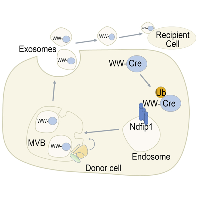
Exosomes are nanovesicles capable of intercellular communication. As such, they represent a highly attractive delivery vehicle. Sterzenbach et al. investigated a mechanism for loading exosomes with exogenous proteins through the sequestration of an evolutionary conserved pathway. Using this method, they loaded exosomes with Cre recombinase and, through nasal administration, have identified the functional delivery of exosomes over large distances into the brain.
Introduction
A critical component for devising drug delivery systems is to package and protect molecules with the ability to release them specifically in a temporal and spatial manner. To date, several systems have been developed as drug delivery vehicles; however, many have limitations with regard to immunogenicity, packaging efficiency, and stability. Interest in vectors has shifted to naturally occurring nanovesicles called exosomes that can package proteins, lipids, DNA, and various forms of RNA for delivery to recipient cells.1 The therapeutic use of exosomes is boosted by their low immunogenicity and toxicity but high biological permeability and biocompatibility.2 These properties enhance exosomes as suitable vehicles for biomolecular delivery and potential avenues for treating a number of diseases.
Exosome are secreted by most cell types and can be found in all bodily fluids.3 The biogenesis of exosomes via multivesicular bodies (MVBs) results in the packaging of both membrane proteins and specific components of the cytoplasm. Most components enriched in exosomes are molecules common to all exosomes irrespective of their cellular origin, indicating a molecular set for core function. In addition, a subset of cellular components is cell-type specific; these components are known to vary with the physiological state of the cell through as-yet poorly defined mechanisms.4
Findings have shown the potential of exosomes to treat neurological diseases;5 conversely, exosomes have been shown to be important in many pathological states.6, 7 Therefore, if exosomes are to be exploited as carriers for therapeutic targeting of diseased tissues, new mechanisms are required for tailoring their cargo. To date, there have been several non-specific methods for loading cargo into exosomes; these include electroporation,8 transfection methods,9 and passive diffusion of components.10 Most of these methods are concerned with loading short RNA sequences,8 with variable degrees of success.11 In contrast, there have been few reports on the successful loading of functional proteins into exosomes.
In the present report, we have exploited a vesicle trafficking mechanism that is conserved from yeast to humans, involving late-domain (L-domain) proteins, ubiquitin, and the Endosomal Sorting Complex Required for Transport (ESCRT) machinery. L-domains are used for the recruitment of ESCRT components, as well as ubiquitin ligases, to cell membranes and are required in the budding of most membrane-enveloped viruses, as well as MVB formation.12 Previously, the L-domain protein syntenin (containing LYPX[n]L motifs) was identified in the recruitment of ALIX (an ESCRT-associated protein) and the subsequent formation and loading of exosomes.13 Similarly, the L-domain-containing protein Ndfip1 (containing PPxY motifs) has been identified in viral budding14 and loading of proteins into exosomes.15, 16 Here we have exploited this pathway to load specific proteins into exosomes. Labeling of an intracellular target protein with a WW tag, which is recognized by the L-domain motifs on Ndfip1, resulted in the loading of the target protein into exosomes. Using this mechanism, we have engineered exosomes containing Cre recombinase that have been used to identify functional delivery of exosomes across the blood-brain barrier to recipient neurons in the brain.
Results
Fusion of a WW Tag to Target Proteins Results in Efficient Loading into Exosomes
In the present study, we investigated a new mechanism to load a specific target protein into exosomes. Previously, we found that Ndfip1, an ubiquitin ligase adaptor protein, was able to export binding partners outside of the cell in exosomes.15, 16 One class of Ndfip1 binding proteins found to be exported in exosomes is the Nedd4 family of ubiquitin ligases.15 Ndfip1 interacts with the WW domains of Nedd4 family ubiquitin ligases through three L-domain motifs (PPxY).17 On this basis, we hypothesized that adding a WW tag onto a target protein would result in interaction of the tagged protein with Ndfip1, leading to ubiquitination and subsequent export in exosomes. To test this hypothesis, we constructed a plasmid coding for a fusion protein containing two WW regions linked to Cre recombinase (WW-Cre). The recombinase provides a functional reporter for exosome-mediated transfer of the protein into cells harboring floxed fluorescent proteins.
To test whether the addition of a WW tag interfered with Cre activity, we analyzed the efficiency of WW-Cre compared to standard Cre for recombinase activity. We first investigated whether the WW tag interfered with Cre localization when transfected into cells and found no change in WW-Cre subcellular localization compared to Cre (Figure S1). Next, mouse embryonic fibroblasts (MEFs) derived from membrane Tomato/membrane GFP (mT/mG) reporter mice were transfected with both control Cre and WW-Cre to detect recombinase activity. Normally, mT/mG MEFs are red, but after Cre recombination, there is a change to green due to excision of the floxed membrane Tomato (mT) cassette and expression of membrane GFP (mG). Flow cytometry was performed to quantitatively analyze the frequency of recombination between the two forms of Cre. The results showed that WW-Cre was as efficient as Cre after transfection of mT/mG MEFs, indicating that there was no adverse effect of the WW tag on the recombinase protein (Figure 1A; dot plots shown in Figure S2A).
Figure 1.

The Packaging of Targeted Fusion Proteins into Exosomes
(A) Flow cytometric analysis of transfected mT/mG MEFs showed that the WW-tagged Cre protein (WW-Cre) maintained Cre-recombinase activity compared to wild-type Cre protein. Unpaired t test. (B) Cre protein is not exported in exosomes with or without Ndfip1 expression. WW-Cre protein is efficiently exported into exosomes in the presence of Ndfip1 in LN18 cells. The asterisk indicates the WW-Cre band above the Tsg101 band after reprobing the western blot. (C) NanoSight tracking analysis was used to measure the particle size of the purified exosomes. (D) GW4869 inhibited the release of WW-Cre protein into exosomes. Data points are the average ± SEM of three independent experiments.
The next set of experiments tested for the possibility of spontaneous Cre protein loading into exosomes without the requirement of Ndfip1 and for the necessity of binding to Ndfip1 before Cre can be exported inside exosomes. In previous studies, expression of Cre in cells has not resulted in Cre protein being exported and detected in exosomes.18, 19 Similarly, we found that transient transfection of Cre into LN18 and HEK293T cells did not result in the transport of Cre protein into exosomes in the presence of either the endogenous or the overexpressed Ndfip1 (Figure 1B, exosome lanes 1 and 2; Figure S2B, exosome lanes 1 and 2). In contrast, the WW-Cre fusion protein can be found in the purified exosomes depending on the level of Ndfip1 present in the cell (Figure 1B, exosome lane 4; Figures S2B and S2C, exosome lane 4), indicating the dependence on Ndfip1 for exosome loading of WW-Cre. To confirm the presence of exosomes, particle size distribution was performed using NanoSight analysis (Figure 1C). A linear sucrose gradient also indicated that the exosome preparation showed the correct density for the fraction, which contained WW-Cre protein and the exosomal marker Tsg101 (Figure S2D). To confirm the trafficking of different WW-tagged proteins into exosomes, we used a WW-mCherry construct. In accordance with the WW-Cre findings in LN18 cells, WW-mCherry was only found in exosomes in the presence of Ndfip1 expression (Figure S2E).
To exclude the function of unspecific release of WW-Cre from the cell, resulting in recombination independent of exosome transfer, we prevented exosome release using the sphingomyelinase inhibitor GW4869.20 Because dying cells release apoptotic bodies that can have a size distribution similar to that of exosomes, we performed cell death assays with different concentrations of GW4869 using phosphatidylinositol (PI) staining combined with flow cytometry. Significant cell death compared to the control was detected at 5 and 10 μM, but not at 2.5 μM, of GW4869 treatment (Figure S3). LN18 cells were co-transfected with both WW-Cre and Ndfip1 and cultured for 2 days with or without 2.5 μM GW4869 before exosomes were purified from the supernatant. In the untreated cells, we observed WW-Cre in the exosome preparation; in contrast, cells treated with GW4869 inhibited the release of WW-Cre into the exosome preparation (Figure 1D). The impaired release of exosomes by GW4869 was confirmed by the reduction of Tsg101 in the exosome fraction. In summary, our results show that a target protein can be labeled with a WW tag, resulting in exosomal export in an Ndfip1-dependent manner.
WW-Cre in Exosomes Is Functionally Active following Uptake by Recipient Cells
The tagged WW-Cre protein in exosomes was then investigated for functional biological activity following uptake in recipient cells. The human glioblastoma cell line LN18 was used as the donor cell for the production of exosomes containing WW-Cre, and the reporter line mT/mG was used as the recipient cell type to monitor the uptake and activity of exosomes containing WW-Cre. In control experiments, LN18 cells were transfected with three varieties of plasmid DNA: Cre, WW-Cre, and Cre plus Ndfip1. mT/mG reporter cells were incubated with purified exosomes for 72 hr to allow for exosome uptake and DNA recombination. The results showed that none of the three conditions resulted in Cre-mediated DNA recombination in mT/mG reporter cells (Figures 2A–2C). In contrast, exosomes derived from LN18 cells transfected with WW-Cre and Ndfip1 showed robust Cre recombination in reporter cells (Figure 2D). Quantification of Cre-mediated recombination in reporter cells showed that only exosomes derived from donor cells with WW-Cre and Ndfip1 transfection reported significant Cre recombinase activity (Figure 2E).
Figure 2.

Exosomes Containing Target WW-Cre Proteins Are Functionally Active in Recipient Reporter Cells
(A–D) Exosomes purified from LN18 cells transfected with Cre only (A), Cre and Ndfip1 (B), WW-Cre only (C), or WW-Cre and Ndfip1 (D) were delivered to mT/mG MEFs. (E) Quantification of the number of positive reporter mT/mG MEFs after exosome delivery. Scale bar, 50 μm. Data points are the average ± SEM of three independent experiments. One-way ANOVA tests with Bonferroni post-tests, ****p < 0.0001.
Ndfip1 Is Required for Transport of WW-Cre into Exosomes
Ndfip1 is an adaptor molecule for the Nedd4 family of ubiquitin ligases and can select target molecules for binding and E3 ligase ubiquitination.17, 21, 22 To investigate WW-Cre binding to Ndfip1, both proteins were expressed in HEK293T cells and immunoblotted for Cre following immunoprecipitation using antibodies to Ndfip1. WW-Cre was only detected when Ndfip1 was co-expressed, indicating the direct interaction of the two proteins. WW-Cre was not pulled down when Ndfip1 was not co-expressed, showing the specificity of the interaction between Ndfip1 and WW-tagged protein (Figure 3A).
Figure 3.

Ndfip1 Expression Is Required for Functional WW-Cre Exosomes and Mediates the Ubiquitination of WW-Cre
(A) Immunoprecipitation assay of overexpressed WW-Cre and Ndfip1 in HEK293T cells showed an interaction between the two proteins. (B) Ndfip1+/+ and Ndfip1−/− MEFs used in the co-culture assay in (C) expressed similar levels of WW-Cre, as shown by western blotting. (C) Ndfip1+/+ and Ndfip1−/− MEFs infected with lentiviral construct of WW-Cre were co-cultured with mT/mG reporter MEFs. Only Ndfip1+/+ MEFs resulted in a positive recombination of mT/mG MEFs. (D) Denaturing ubiquitin assays show that Cre protein is not ubiquitinated with or without Ndfip1. In contrast, WW-Cre protein is predominantly monoubiquitinated in the presence of Ndfip1. Scale bar, 50 μm.
To further investigate the requirement of Ndfip1 for exosomal transport of WW-tagged proteins, we performed functional assays using MEFs derived from Ndfip1 knockout mice (Figure 3B). Because these assays used endogenous levels of Ndfip1 and exosome production, we performed the experiments using co-culture assays (to increase the sensitivity of the assay) with both Ndfip1+/+ and Ndfip1−/− MEFs as the donor cells. Both Ndfip1+/+ and Ndfip1−/− MEFs were infected with a lentivirus containing the WW-Cre construct (Figure 3B) 24 hr before being washed and placed in co-culture with the reporter mT/mG cells. Analysis of cells after 72 hr in co-culture showed that only Ndfip1+/+ MEFs were able to promote recombination in mT/mG MEFs; no recombination was observed in Ndfip1−/− MEF co-cultures (Figure 3C). These experiments reveal that endogenous Ndfip1 in MEFs was required for the transfer of WW-Cre in exosomes, leading to recombination in co-cultured reporter cells.
Given that Ndfip1 is closely linked to the ubiquitination of target proteins, we next investigated the role of Ndfip1 in mediating the ubiquitination of both Cre and WW-Cre using HEK293T cells and denaturing ubiquitination assays. Expression of Cre protein alone or in the presence of Ndfip1 did not result in ubiquitination of Cre (Figure 3D, lanes 2 and 3). Expression of WW-Cre alone resulted in limited ubiquitination, most likely due to low levels of endogenous Ndfip1 in HEK293T cells (Figure 3D, lane 4). Co-expression of WW-Cre and Ndfip1 resulted in efficient ubiquitination of WW-Cre, with the predominant ubiquitination form observed to be monoubiquitination (Figure 3D, lane 5). These results suggest a mechanism in which Ndfip1 directly interacts with and mediates the ubiquitination of WW-Cre, resulting in exosomal transport of the protein.
WW-Cre Protein Is Localized inside Exosomes
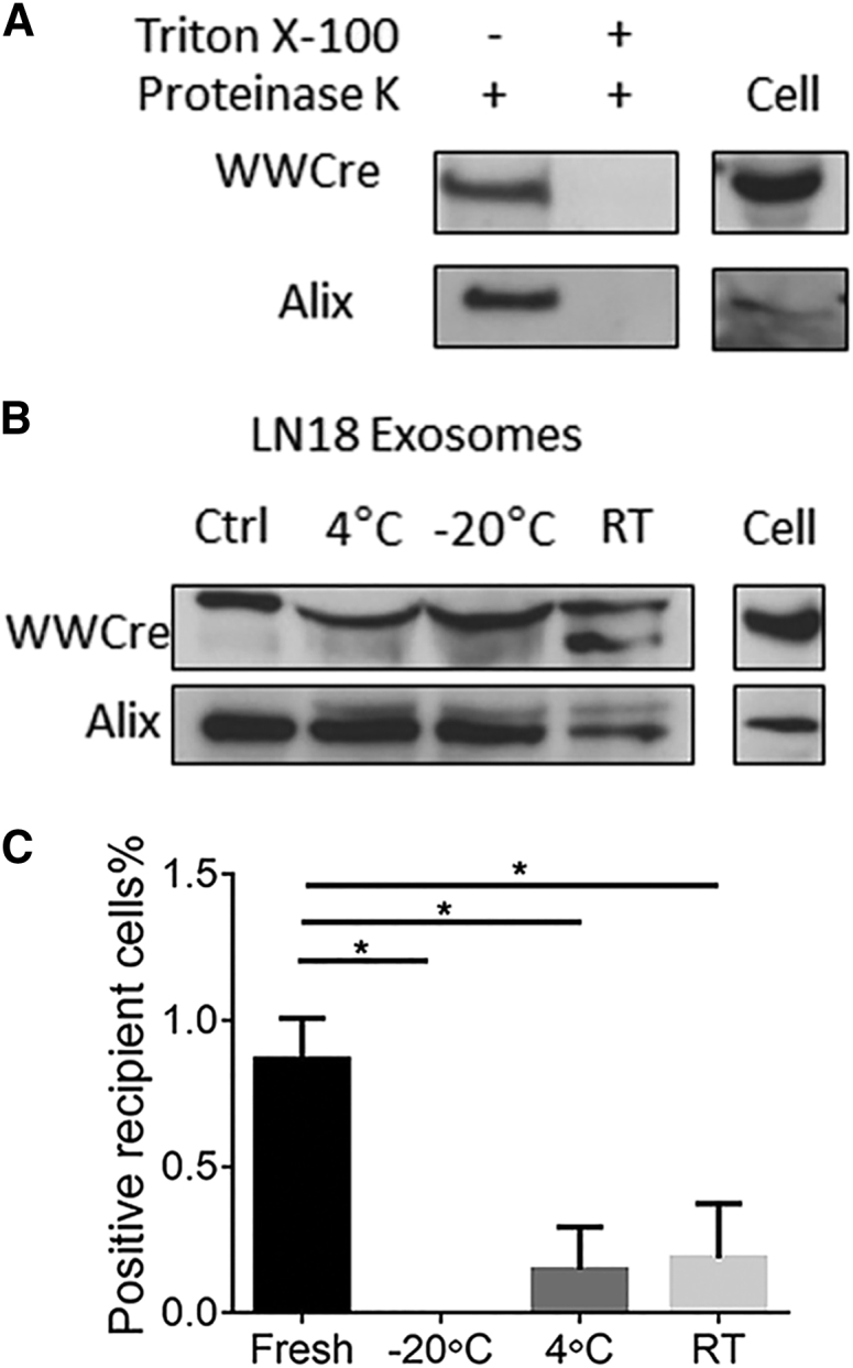
Exosomes have been shown to promote the transport of proteins both within and on the exterior of the vesicle, with proteins such as α-synuclein observed to be transported in both environments.23 Transport on the exterior of exosomes does not provide an environment to protect proteins from extracellular proteases and would therefore not be suitable for therapeutic applications. To investigate the exosomal location of WW-tagged proteins, we performed protease digestion assays. Exosomes purified from LN18 cells were treated with proteinase K in the presence or absence of Triton X-100. Proteinase K treatment without Triton X-100 did not result in the degradation of WW-Cre protein, as shown by western blotting, indicating that the tagged protein is contained and protected within the exosome microenvironment (Figure 4A). In contrast, exosomes treated with Triton X-100 and proteinase K showed a loss of WW-Cre protein and the exosome marker Alix (Figure 4A). These results indicate that the WW-tagged Cre protein is localized inside exosomes when secreted from the cell.
Figure 4.
The Stability and Function of Stored Exosomes
(A) Proteinase treatment of exosomes did not result in the degradation of WW-Cre unless Triton X-100 was added to permeabilize the exosome membrane. (B) Exosomes were stored for 3 days under different conditions before western blotting was performed to determine the stability of WW-Cre protein compared to a control fresh preparation of WW-Cre exosomes. (C) Stored exosomes were analyzed for the ability to deliver active WW-Cre to mT/mG reporter MEFs. Data points are the average ± SEM of three independent experiments. One-way ANOVA tests with Bonferroni post-tests, *p < 0.05.
Exosomes with WW-Cre Provide a Functional Assay for Exosome Storage
Previous reports have questioned the stability of exosomes under various storage conditions, particularly for long periods.24, 25 These reports rely on western blots to identify protein degradation products but fail to provide additional information on their functional integrity after exosome uptake in recipient cells. The WW-Cre system affords a functional assay to assess the biological activity of exosomes following storage. Western blot analysis showed that WW-Cre protein was stable in exosomes stored below 4°C over 3 days (Figure 4B), similar to other reported findings.25 However, when tested for biological activity using the mT/mG recombinase assay, the stored exosomes reported reduced functional Cre activity compared to fresh preparations of exosomes (Figure 4C). Because the WW-Cre protein was not degraded over these periods (as observed in western blots) (Figure 4B), these findings suggest that following storage, exosomes are compromised in their ability to transfer proteins into recipient cells. Alternatively, the exosomes have increased fragility following storage, rendering them and their contents susceptible to degradation.
Nasal Delivery of Exosomes with WW-Cre into the Brain
Exosomal delivery of specific biomolecules in vivo has predominantly involved the transfer of exosomal RNA between cells.8 It remains to be seen whether proteins inside exosomes can also be delivered into the brain with intact biological activity. To test this, exosomes containing WW-Cre were used to test for delivery and recombination inside the brains of transgenic mice carrying the loxP stop cassette linked to tdTomato expression.26 LN18 glioblastoma cells were chosen as the donors for harvesting exosomes due to their reliability in generating biologically active WW-Cre in exosomes in vitro. Control exosomes were harvested from non-transfected LN18 cells, as well as from cells transfected with Cre (not WW-Cre) and Ndfip1. These different categories of exosomes were then delivered to the reporter mouse strain Ai14 using the nasal route previously shown to effectively target exosomes to the brain.27 Exosomes were delivered at post-natal day (P) 7, P10, and P13 before analysis at P28 for tdTomato expression in brain sections. Analysis of control brains for tdTomato expression showed that a small number of cells were able to undergo spontaneous recombination independent of any Cre protein, with exosomes derived from non-transfected LN18 cells, as well as Cre- and Ndfip1-transfected cells showing a low level of recombination in these reporter mice (Figure 5A). By contrast, brains exposed to purified exosomes from cells transfected with both WW-Cre and Ndfip1 showed a significantly increased number of recombined cells (Figures 5A–5C). Cells with positive signals were dispersed throughout the brain, including the olfactory bulb, cortex, striatum, hippocampus, and cerebellum (Figure 5C; distribution of positive cells throughout the brain shown in Figure S4). Counterstaining for neuronal-specific nuclear protein (NeuN) immunoreactivity confirmed the neuronal nature of recipient cells (Figure 5D). In addition, a limited number of microglia (identified by ionized calcium binding adaptor molecule 1 [Iba-1] staining) demonstrated Cre-mediated recombination (Figure 5E). We conclude that nasal delivery of exosomes allows for the transport of functional target proteins to multiple brain regions.
Figure 5.

In Vivo Activity of Exogenous Exosomes
(A) Quantification of positive reporter cells after nasal delivery of exosomes derived from LN18 cells transfected with WW-Cre and Ndfip1, Cre and Ndfip1, or untransfected to Ai14 mice. Mouse brains were collected 14 days after last nasal delivery, and sections from multiple brain regions were analyzed for red reporter cells indicating Cre-recombination (n = 4 mice per condition). (B) Overview of the coronal brain section analyzed for Cre recombination, with the white box indicating the area imaged in (C). (C) Higher magnification of brain regions showing Cre recombination after delivery of WW-Cre containing exosomes. (D) Immunocytochemistry with anti-NeuN antibody shows colocalization in cells with Cre-recombinase activity (tdTomato) in Ai14 mice after WW-Cre exosome delivery. (E) Immunocytochemistry with anti-Iba-1 antibody shows colocalization with Cre-recombinase activity (tdTomato) in Ai14 mice after WW-Cre exosome delivery. One-way ANOVA tests with Bonferroni post-tests, *p < 0.05. Scale bars, (B) 2.5 mm, (C) 250 μm, and (D and E) 50 μm.
Discussion
Exosomes directly and indirectly control a diverse range of biological processes through the transport of signaling molecules such as proteins and nucleic acids.28 The ability to act as an in vivo transport vehicle, protecting cargo from the extracellular environment, has resulted in the identification of exosomes as both effectors and biomarkers of disease processes.29 In addition, exosomes have been promoted as cell-free vectors to deliver molecules for therapeutic modification of diseased tissues.2 What has been lacking thus far is a mechanism to load proteins of interest into exosomes. To address this, we have developed a system to specifically package exogenous proteins into exosomes and have used it to deliver functional protein to the brain.
To develop a specific mechanism for the packaging of proteins into exosomes, we investigated a pathway known to be involved in membrane budding and formation of MVBs. Proteins containing L-domains (so called because they were initially identified to function late in the viral budding process) are found in retroviruses, rhabdoviruses, and filoviruses, as well as cellular proteins involved in endosome transport, and have an important function in triggering vesicle formation. Because the formation of exosomes requires the intraluminal budding of endosomal membranes, it is not surprising that L-domain proteins such as syntenin have been identified to interact with ESCRT components, resulting in the sorting and biogenesis of exosomes.13 Ndfip1 contains three highly conserved L-domain motifs known to interact with the Nedd4 ubiquitin ligase family.17 We have previously shown that Ndfip1 is involved in the trafficking of proteins on Rab5 early endosomes using the ubiquitin pathway,30 and for a number of Ndfip1-interacting proteins, this can result in packaging into exosomes.15, 16 Here we show that attaching a WW tag onto a target protein results in a direct interaction with Ndfip1, leading to monoubiquitination of the target protein and exosomal packaging. Monoubiquitination has previously been shown to be required for ESCRT machinery recognition used in the formation of MVBs.31 Thus, Ndfip1 expression can act as a molecular switch for the transport of WW-tagged proteins into exosomes.
The WW-Cre exosome system has potential to serve as a tool to answer questions in the field regarding the nature of exosome biocompatibility between cells. For instance, are exosomes from one cell type more likely to communicate with homologous compared to heterologous cells? Is there an exosome code of receptors or ligands on the surface of exosomes that are produced and recognized by different cell types? Given the vast nature of cell types in the body that can produce exosomes, it is unclear how the diverse origins of exosomes (and cargo) and equally diverse repertoire of potential recipient cells are able to maintain biological order. Here we identify homologous communication pathways in vitro between MEFs, as well as heterologous communication in vivo between glioblastoma cells and neurons. This potential for specific delivery of exosomes to organs and cells types should be further investigated and exploited for targeted delivery of biomolecules via exosomes. By the same token, caution should be used when interpreting the role of exosomes using in vitro assays, because negative findings may represent a lack of communication between exosomes and recipient cells, rather than a deficiency of exosome function.
Engineering exosomes with a reporter function also opened up the opportunity to investigate the functional activity of stored exosomes. Investigating the stability of exosomes is an important aspect if they are to be used in therapeutic settings. There is no consensus about the ability to store exosomes without affecting the biological activity of the protein contents. Some reports have indicated the successful storage of exosomes for months or years;24 in contrast, other reports showed that short-term storage (72 hr) at 37°C and 4°C led to aggregation of exosomes with changes in exosome size.32 Regardless, most of these assays have relied on the extent of protein degradation as gauged by western blot analysis, rather than an assay for functional protein activity after delivery. We observed that exosomes stored under multiple conditions for 72 hr resulted in a loss of functional activity in recipient cells. Because the exosomal WW-Cre protein was not degraded after storage (as observed by western blotting), our data suggest that either there is a loss of exosomal surface proteins that are required for recipient cell recognition or damage has occurred to the exosome during storage, resulting in degradation preventing entry in the recipient cell. Further studies investigating methods to maintain exosome function after storage, such as using protease inhibitors in the storage conditions, may enhance the ability to store functional exosomes long term.
In the current experiments, the nasal route was chosen as a non-invasive method to deliver exosomes containing exogenous protein to the brain. It has previously been reported that exosomes can cross the blood-brain barrier to deliver therapeutics, using dyes as a proxy to track exosome location.27 However, the use of membrane-bound dyes does not identify whether exosomes can deliver functional cargo and dye labeling does not always faithfully report the location of exosomes, because the dye can diffuse in vivo and stick to the surface of cells and vessels. Using WW-Cre reporter exosomes, we have shown that nasal delivery results in the transport of biologically active proteins across the blood-brain barrier to a number of brain regions. Our findings do not identify the translocation pathway for exosomes from the nasal cavity to the brain, but our results are consistent with trafficking along perineuronal and/or perivascular channels,27 given the distribution observed and the lack of sequential labeling of cells required for intracellular transport.33 These glioma-derived exosomes were mainly taken up by neurons in the brain, with a smaller number of positive microglia also observed. No astrocytes or oligodendrocytes were found to uptake exogenous exosomes derived from glia. These results are consistent with reports showing that glial cells can communicate with neurons in vivo through exosome transmission.34 This is in contrast to exosomes derived from T cells that have been shown to predominantly communicate with microglia when delivered to the brain.27 Altogether, these findings support the notion that exosomes derived from different cell linages have preferential recipient cell types.
Research in animal models has shown the potential of exosomes to treat disease,35 and the first clinical trials using exosomes are being undertaken.36 However, our current understanding of exosome trafficking, function, and stability is poorly characterized. Here we have appropriated an evolutionary conserved pathway used to secrete molecules from the cell to create a system that can specifically load proteins into functionally active exosomes. Using this system, we have investigated exosomal target protein delivery both in vitro and in vivo. The ability to engineer these nature-based delivery vehicles for the transmission of protein cargo across the blood-brain barrier represents an important step in the development of exosome-based therapeutics.
Materials and Methods
Cell Culture
A stable mT/mG reporter MEF line was produced from the global double-fluorescent Cre reporter mouse line mT/mG.37 mT/mG MEFs were cultured from an embryonic day (E) 13.5 embryo and infected with a lentivirus contain the SV40 large T antigen.38 Ndfip1+/+ and Ndfip1−/− MEFs have previously been described.39 All cells, including HEK293T, MEFs, and LN18 glioblastoma cells, were cultured in DMEM containing 10% fetal calf serum, 50 units of penicillin, 50 μg/mL streptomycin, and 4 mM L-glutamine (Invitrogen) and were maintained at 37°C with 5% CO2. Transfection of plasmids was performed using TurboFect Transfection Reagent (Thermo Scientific) according to the manufacturer’s instructions.
Plasmids and Flow Cytometry
FLAG-tagged Ndfip1 (FLAG-Ndfip1) in pcDNA317 and His-ubiquitin in pcDNA340 have been previously described. WW-Cre and WW-mCherry in pcDNA3, as well as a WW-Cre lentivirus, were constructed by cloning WW domains 2 and 3 of Nedd4-1 to the N terminus of Cre. The efficiency of the WW-Cre construct for Cre activity was analyzed through comparison with a control Cre expression vector using flow cytometry. Briefly, mT/mG MEFs were transiently transfected using TurboFect Transfection Reagent (Thermo Scientific) with Cre or WW-Cre and cultured for 72 hr. Cells were trypsinized, washed in PBS, and then resuspended in 200 μL PBS before being analyzed using a BD FACSCaliber flow cytometer. Each analysis included 100,000 cells, with a color change from green to red indicating positive recombination.
Isolation of Exosomes and Sucrose Gradient
Cells were cultured for up to 72 hr post-transfection before supernatant was collected. The supernatant was cleared from dead cells and debris by centrifugation for 10 min at 200 g followed by centrifugation for 20 min at 20,000 g. Exosomes were isolated from the supernatant by centrifugation for 70 min at 100,000 g and washed with ice-cold PBS before being centrifuged again for 70 min at 100,000 g. Exosomes used for western blotting were resuspended in 30 μL of radioimmunoprecipitation assay (RIPA) buffer (50 mM Tris [pH 8], 100 mM NaCl, 5 mM EDTA, 1% Triton X-100, 0.2% SDS, protease inhibitor cocktail [PIC]) and boiled after the addition of 10 μL Laemmli buffer before application. Exosomes used for uptake or intranasal delivery were resuspended in HEPES buffer at a protein concentration of ∼2 μg/μL. For sucrose-gradient experiments, pelleted exosomes were resuspended in 1 mL of 0.25 M sucrose and PBS before being loaded on top of a linear sucrose gradient (0.25 to 2.5 M). The gradient was centrifuged at 100,000 g for 16 hr before single fractions were collected, diluted in ice-cold PBS, centrifuged at 100,000 g for 70 min, and prepared for western blotting.
Particle Size Analysis
Exosomes were purified from LN18 cells and diluted 50% in PBS buffer. Real-time high-resolution particle detection, counting, and sizing were performed on the NanoSight NS300 following manufacturer protocols (Malvern Instruments). Particle concentration (in particles per milliliter) and size was calculated by the NanoSight system.
Co-culture and Purified Exosome Uptake Assays
mT/mG MEFs were cultured on coverslips in a 12-well plate for 24 hr at 37°C with 5% CO2 before Ndfip1+/+ or Ndfip1−/− MEFs infected with WW-Cre were added and cultured under the same conditions for 72 hr. Each well was washed with ice-cold PBS before being fixed in methanol for 10 min at −20°C. After fixation, coverslips were washed three times with ice-cold PBS; the second wash also contained DAPI for nuclear staining. For exosome uptake assays, exosomes were purified via ultracentrifugation and the exosome pellet was resuspended in HEPES buffer. Exosomes were either directly added after purification (fresh exosomes) or stored in HEPES buffer at −20°C, 4°C, or room temperature (∼21°C) before being added to mT/mG MEFs (250 μg of protein vesicles by Bradford measurement were added in vitro). Cells were cultured for 72 hr after the addition of exosomes to allow for uptake and recombination before coverslip mounting for microscopy.
Immunoprecipitation Assay and Western Blotting
10-cm dishes containing 2 × 10⁶ HEK293T cells were transfected either with WW-Cre and an empty pcDNA3 plasmid as a control or with WW-Cre and Ndfip1-Flag using TurboFect Transfection Reagent (Thermo Scientific). Cell lysates were prepared 24 hr post-transfection before immunoprecipitation assays were performed with anti-FLAG M2 Affinity Gel according to the manufacturer’s instructions (Sigma-Aldrich). Ubiquitination assay was performed using HisLink beads (Promega) under denaturing conditions using 6 M guanidine HCl/RIPA containing N-ethylmaleimide to inhibit deubiquitinase enzyme activity. After washing the beads three times in guanidine-HCl/RIPA, the fractions were eluted in 300 mM imidazole and boiled after the addition of Laemmli buffer. Cell lysates and isolated exosomes were subjected to 10% SDS-PAGE and western blotting according to standard protocols. Western blots were incubated at 4°C for 16 hr with the indicated primary antibodies and washed three times in Tris-buffered saline T (TBS-T), followed by 1 hr of incubation in secondary antibody at room temperature. Proteins were detected using enhanced chemiluminescence (ECL) reagent according to the manufacturer’s instructions (GE Healthcare) and visualized by exposure to X-ray film.
Intranasal Delivery of Exosomes
All animal procedures were approved by the Florey Institute of Neuroscience and Mental Health Animal Ethics Committee. Ai14 mice were used as a reporter line26 for intranasal delivery of exosomes. LN18 cells, either transfected with WW-Cre and Ndfip1 (WW-Cre + Ndfip1) or Cre and Ndfip1 (Cre+Ndfip1) or as untransfected control (Ctrl), were cultured for 72 hr before exosomes were purified and resuspended in HEPES buffer (protein concentration of 2 μg/μL). Purified exosomes were administered at P7, P10, and P13 to awake mice. The intranasal grip was used to immobilize the mice.41 A total of 6 μL was administered intranasally as drops with a small micropipette to each side of the nasal cavity per time point (meaning a total of 12 μL per time point, resulting in a total delivery of 72 μg of exosomal protein to each mouse). At P28, mice were transcardially perfused with PBS, followed by a 4% paraformaldehyde solution (pH 7.4). Brain tissue was post-fixed for 2 hr in 4% paraformaldehyde and then cryopreserved in PBS containing 30% sucrose. Brains were embedded in optimal cutting temperature compound (OCT) (Tissue-Tek, Sakura) and kept at −80°C. Brain tissue sections were cut on the coronal plane with a cryostat (14 μm thick), and the tissue sections were stored at −80°C. Analysis for recombined cells was performed on 36 sections per brain, combined with staining for either NeuN or Iba-1. Six sections in six areas (Bregma 4.5, 2.0, 0, −1.5, −2.5, and −5.5), covering areas of the olfactory bulb, cortex, striatum, hippocampus, and cerebellum, were analyzed for recombination.
Antibodies and Chemicals
Antibodies used in this study were rat monoclonal anti-Ndfip1,39 mouse anti-GM130 antibody (BD Biosciences), rabbit anti-Tsg101 (Sigma-Aldrich), rabbit anti-Cre (BioLegend), mouse anti-FLAG M2 antibody (Sigma-Aldrich), mouse anti-Alix (Cell Signaling), mouse monoclonal antibody to NeuN (Chemicon), and rabbit polyclonal antibody anti-Iba-1 (Wako). Secondary antibodies were Alexa Fluor 488-conjugated goat anti-rabbit immunoglobulin G (IgG) (1:500; Molecular Probes), and Alex 488-conjugated goat anti-mouse IgG (1:500; Invitrogen). The horseradish peroxidase (HRP)-conjugated secondary antibodies used were goat polyclonal anti-rabbit (1:10,000; Millipore), goat polyclonal anti-rat (1:10,000; Millipore), and goat polyclonal anti-mouse (1:15,000; Millipore). The reagents used were GW4869 (Sigma), to inhibit exosome release; DAPI (1:10,000; Dako), for nuclear staining; proteinase K (Sigma-Aldrich); Triton X-100 (MP Biomedicals); and trypsin-EDTA (0.05%; Thermo Fisher).
Author Contributions
U.S., U.P., L.-H.L., and J.H. performed research and analyzed data. U.S., J.S., S.-S.T., and J.H. designed the research and wrote the manuscript.
Conflicts of Interest
All authors declare no competing financial interests or conflicts.
Acknowledgments
This work was supported by the Australia National Health and Medical Research Council through project grants (1066925 and 1066895) and the Victorian Government through the Operational Infrastructure Scheme.
Footnotes
Supplemental Information includes four figures and can be found with this article online at http://dx.doi.org/10.1016/j.ymthe.2017.03.030.
Supplemental Information
References
- 1.Simons M., Raposo G. Exosomes—vesicular carriers for intercellular communication. Curr. Opin. Cell Biol. 2009;21:575–581. doi: 10.1016/j.ceb.2009.03.007. [DOI] [PubMed] [Google Scholar]
- 2.EL Andaloussi S., Mäger I., Breakefield X.O., Wood M.J. Extracellular vesicles: biology and emerging therapeutic opportunities. Nat. Rev. Drug Discov. 2013;12:347–357. doi: 10.1038/nrd3978. [DOI] [PubMed] [Google Scholar]
- 3.Vlassov A.V., Magdaleno S., Setterquist R., Conrad R. Exosomes: current knowledge of their composition, biological functions, and diagnostic and therapeutic potentials. Biochim. Biophys. Acta. 2012;1820:940–948. doi: 10.1016/j.bbagen.2012.03.017. [DOI] [PubMed] [Google Scholar]
- 4.Mathivanan S., Lim J.W., Tauro B.J., Ji H., Moritz R.L., Simpson R.J. Proteomics analysis of A33 immunoaffinity-purified exosomes released from the human colon tumor cell line LIM1215 reveals a tissue-specific protein signature. Mol. Cell. Proteomics. 2010;9:197–208. doi: 10.1074/mcp.M900152-MCP200. [DOI] [PMC free article] [PubMed] [Google Scholar]
- 5.Zhang Y., Chopp M., Meng Y., Katakowski M., Xin H., Mahmood A., Xiong Y. Effect of exosomes derived from multipluripotent mesenchymal stromal cells on functional recovery and neurovascular plasticity in rats after traumatic brain injury. J. Neurosurg. 2015;122:856–867. doi: 10.3171/2014.11.JNS14770. [DOI] [PMC free article] [PubMed] [Google Scholar]
- 6.Iero M., Valenti R., Huber V., Filipazzi P., Parmiani G., Fais S., Rivoltini L. Tumour-released exosomes and their implications in cancer immunity. Cell Death Differ. 2008;15:80–88. doi: 10.1038/sj.cdd.4402237. [DOI] [PubMed] [Google Scholar]
- 7.Yang C., Robbins P.D. The roles of tumor-derived exosomes in cancer pathogenesis. Clin. Dev. Immunol. 2011;2011:842849. doi: 10.1155/2011/842849. [DOI] [PMC free article] [PubMed] [Google Scholar]
- 8.Alvarez-Erviti L., Seow Y., Yin H., Betts C., Lakhal S., Wood M.J. Delivery of siRNA to the mouse brain by systemic injection of targeted exosomes. Nat. Biotechnol. 2011;29:341–345. doi: 10.1038/nbt.1807. [DOI] [PubMed] [Google Scholar]
- 9.Mizrak A., Bolukbasi M.F., Ozdener G.B., Brenner G.J., Madlener S., Erkan E.P., Ströbel T., Breakefield X.O., Saydam O. Genetically engineered microvesicles carrying suicide mRNA/protein inhibit schwannoma tumor growth. Mol. Ther. 2013;21:101–108. doi: 10.1038/mt.2012.161. [DOI] [PMC free article] [PubMed] [Google Scholar]
- 10.Fuhrmann G., Serio A., Mazo M., Nair R., Stevens M.M. Active loading into extracellular vesicles significantly improves the cellular uptake and photodynamic effect of porphyrins. J. Control. Release. 2015;205:35–44. doi: 10.1016/j.jconrel.2014.11.029. [DOI] [PubMed] [Google Scholar]
- 11.Ohno S., Takanashi M., Sudo K., Ueda S., Ishikawa A., Matsuyama N., Fujita K., Mizutani T., Ohgi T., Ochiya T. Systemically injected exosomes targeted to EGFR deliver antitumor microRNA to breast cancer cells. Mol.. Ther. 2013;21:185–191. doi: 10.1038/mt.2012.180. [DOI] [PMC free article] [PubMed] [Google Scholar]
- 12.Freed E.O. Viral late domains. J. Virol. 2002;76:4679–4687. doi: 10.1128/JVI.76.10.4679-4687.2002. [DOI] [PMC free article] [PubMed] [Google Scholar]
- 13.Baietti M.F., Zhang Z., Mortier E., Melchior A., Degeest G., Geeraerts A., Ivarsson Y., Depoortere F., Coomans C., Vermeiren E. Syndecan-syntenin-ALIX regulates the biogenesis of exosomes. Nat. Cell Biol. 2012;14:677–685. doi: 10.1038/ncb2502. [DOI] [PubMed] [Google Scholar]
- 14.König R., Zhou Y., Elleder D., Diamond T.L., Bonamy G.M., Irelan J.T., Chiang C.Y., Tu B.P., De Jesus P.D., Lilley C.E. Global analysis of host-pathogen interactions that regulate early-stage HIV-1 replication. Cell. 2008;135:49–60. doi: 10.1016/j.cell.2008.07.032. [DOI] [PMC free article] [PubMed] [Google Scholar]
- 15.Putz U., Howitt J., Lackovic J., Foot N., Kumar S., Silke J., Tan S.S. Nedd4 family-interacting protein 1 (Ndfip1) is required for the exosomal secretion of Nedd4 family proteins. J. Biol. Chem. 2008;283:32621–32627. doi: 10.1074/jbc.M804120200. [DOI] [PubMed] [Google Scholar]
- 16.Putz U., Howitt J., Doan A., Goh C.P., Low L.H., Silke J., Tan S.S. The tumor suppressor PTEN is exported in exosomes and has phosphatase activity in recipient cells. Sci. Signal. 2012;5:ra70. doi: 10.1126/scisignal.2003084. [DOI] [PubMed] [Google Scholar]
- 17.Harvey K.F., Shearwin-Whyatt L.M., Fotia A., Parton R.G., Kumar S. N4WBP5, a potential target for ubiquitination by the Nedd4 family of proteins, is a novel Golgi-associated protein. J. Biol. Chem. 2002;277:9307–9317. doi: 10.1074/jbc.M110443200. [DOI] [PubMed] [Google Scholar]
- 18.Zomer A., Maynard C., Verweij F.J., Kamermans A., Schäfer R., Beerling E., Schiffelers R.M., de Wit E., Berenguer J., Ellenbroek S.I. In vivo imaging reveals extracellular vesicle-mediated phenocopying of metastatic behavior. Cell. 2015;161:1046–1057. doi: 10.1016/j.cell.2015.04.042. [DOI] [PMC free article] [PubMed] [Google Scholar]
- 19.Ridder K., Keller S., Dams M., Rupp A.K., Schlaudraff J., Del Turco D., Starmann J., Macas J., Karpova D., Devraj K. Extracellular vesicle-mediated transfer of genetic information between the hematopoietic system and the brain in response to inflammation. PLoS Biol. 2014;12:e1001874. doi: 10.1371/journal.pbio.1001874. [DOI] [PMC free article] [PubMed] [Google Scholar]
- 20.Trajkovic K., Hsu C., Chiantia S., Rajendran L., Wenzel D., Wieland F., Schwille P., Brügger B., Simons M. Ceramide triggers budding of exosome vesicles into multivesicular endosomes. Science. 2008;319:1244–1247. doi: 10.1126/science.1153124. [DOI] [PubMed] [Google Scholar]
- 21.Mund T., Pelham H.R. Control of the activity of WW-HECT domain E3 ubiquitin ligases by NDFIP proteins. EMBO Rep. 2009;10:501–507. doi: 10.1038/embor.2009.30. [DOI] [PMC free article] [PubMed] [Google Scholar]
- 22.Sang Q., Kim M.H., Kumar S., Bye N., Morganti-Kossman M.C., Gunnersen J., Fuller S., Howitt J., Hyde L., Beissbarth T. Nedd4-WW domain-binding protein 5 (Ndfip1) is associated with neuronal survival after acute cortical brain injury. J. Neurosci. 2006;26:7234–7244. doi: 10.1523/JNEUROSCI.1398-06.2006. [DOI] [PMC free article] [PubMed] [Google Scholar]
- 23.Emmanouilidou E., Melachroinou K., Roumeliotis T., Garbis S.D., Ntzouni M., Margaritis L.H., Stefanis L., Vekrellis K. Cell-produced alpha-synuclein is secreted in a calcium-dependent manner by exosomes and impacts neuronal survival. J. Neurosci. 2010;30:6838–6851. doi: 10.1523/JNEUROSCI.5699-09.2010. [DOI] [PMC free article] [PubMed] [Google Scholar]
- 24.Ge Q., Zhou Y., Lu J., Bai Y., Xie X., Lu Z. miRNA in plasma exosome is stable under different storage conditions. Molecules. 2014;19:1568–1575. doi: 10.3390/molecules19021568. [DOI] [PMC free article] [PubMed] [Google Scholar]
- 25.Kalra H., Adda C.G., Liem M., Ang C.S., Mechler A., Simpson R.J., Hulett M.D., Mathivanan S. Comparative proteomics evaluation of plasma exosome isolation techniques and assessment of the stability of exosomes in normal human blood plasma. Proteomics. 2013;13:3354–3364. doi: 10.1002/pmic.201300282. [DOI] [PubMed] [Google Scholar]
- 26.Madisen L., Zwingman T.A., Sunkin S.M., Oh S.W., Zariwala H.A., Gu H., Ng L.L., Palmiter R.D., Hawrylycz M.J., Jones A.R. A robust and high-throughput Cre reporting and characterization system for the whole mouse brain. Nat. Neurosci. 2010;13:133–140. doi: 10.1038/nn.2467. [DOI] [PMC free article] [PubMed] [Google Scholar]
- 27.Zhuang X., Xiang X., Grizzle W., Sun D., Zhang S., Axtell R.C., Ju S., Mu J., Zhang L., Steinman L. Treatment of brain inflammatory diseases by delivering exosome encapsulated anti-inflammatory drugs from the nasal region to the brain. Mol. Ther. 2011;19:1769–1779. doi: 10.1038/mt.2011.164. [DOI] [PMC free article] [PubMed] [Google Scholar]
- 28.Colombo M., Raposo G., Théry C. Biogenesis, secretion, and intercellular interactions of exosomes and other extracellular vesicles. Annu. Rev. Cell Dev. Biol. 2014;30:255–289. doi: 10.1146/annurev-cellbio-101512-122326. [DOI] [PubMed] [Google Scholar]
- 29.Fais S., O’Driscoll L., Borras F.E., Buzas E., Camussi G., Cappello F., Carvalho J., Cordeiro da Silva A., Del Portillo H., El Andaloussi S. Evidence-based clinical use of nanoscale extracellular vesicles in nanomedicine. ACS Nano. 2016;10:3886–3899. doi: 10.1021/acsnano.5b08015. [DOI] [PubMed] [Google Scholar]
- 30.Li Y., Low L.H., Putz U., Goh C.P., Tan S.S., Howitt J. Rab5 and Ndfip1 are involved in Pten ubiquitination and nuclear trafficking. Traffic. 2014;15:749–761. doi: 10.1111/tra.12175. [DOI] [PubMed] [Google Scholar]
- 31.Raiborg C., Rusten T.E., Stenmark H. Protein sorting into multivesicular endosomes. Curr. Opin. Cell Biol. 2003;15:446–455. doi: 10.1016/s0955-0674(03)00080-2. [DOI] [PubMed] [Google Scholar]
- 32.Sokolova V., Ludwig A.K., Hornung S., Rotan O., Horn P.A., Epple M., Giebel B. Characterisation of exosomes derived from human cells by nanoparticle tracking analysis and scanning electron microscopy. Colloids Surf. B Biointerfaces. 2011;87:146–150. doi: 10.1016/j.colsurfb.2011.05.013. [DOI] [PubMed] [Google Scholar]
- 33.Thorne R.G., Emory C.R., Ala T.A., Frey W.H., 2nd Quantitative analysis of the olfactory pathway for drug delivery to the brain. Brain Res. 1995;692:278–282. doi: 10.1016/0006-8993(95)00637-6. [DOI] [PubMed] [Google Scholar]
- 34.Frühbeis C., Fröhlich D., Kuo W.P., Amphornrat J., Thilemann S., Saab A.S., Kirchhoff F., Möbius W., Goebbels S., Nave K.A. Neurotransmitter-triggered transfer of exosomes mediates oligodendrocyte-neuron communication. PLoS Biol. 2013;11:e1001604. doi: 10.1371/journal.pbio.1001604. [DOI] [PMC free article] [PubMed] [Google Scholar]
- 35.Rahman M.J., Regn D., Bashratyan R., Dai Y.D. Exosomes released by islet-derived mesenchymal stem cells trigger autoimmune responses in NOD mice. Diabetes. 2014;63:1008–1020. doi: 10.2337/db13-0859. [DOI] [PMC free article] [PubMed] [Google Scholar]
- 36.Morse M.A., Garst J., Osada T., Khan S., Hobeika A., Clay T.M., Valente N., Shreeniwas R., Sutton M.A., Delcayre A. A phase I study of dexosome immunotherapy in patients with advanced non-small cell lung cancer. J. Transl. Med. 2005;3:9. doi: 10.1186/1479-5876-3-9. [DOI] [PMC free article] [PubMed] [Google Scholar]
- 37.Muzumdar M.D., Tasic B., Miyamichi K., Li L., Luo L. A global double-fluorescent Cre reporter mouse. Genesis. 2007;45:593–605. doi: 10.1002/dvg.20335. [DOI] [PubMed] [Google Scholar]
- 38.Ahuja D., Sáenz-Robles M.T., Pipas J.M. SV40 large T antigen targets multiple cellular pathways to elicit cellular transformation. Oncogene. 2005;24:7729–7745. doi: 10.1038/sj.onc.1209046. [DOI] [PubMed] [Google Scholar]
- 39.Howitt J., Lackovic J., Low L.H., Naguib A., Macintyre A., Goh C.P., Callaway J.K., Hammond V., Thomas T., Dixon M. Ndfip1 regulates nuclear Pten import in vivo to promote neuronal survival following cerebral ischemia. J. Cell Biol. 2012;196:29–36. doi: 10.1083/jcb.201105009. [DOI] [PMC free article] [PubMed] [Google Scholar]
- 40.Low L.H., Chow Y.L., Li Y., Goh C.P., Putz U., Silke J., Ouchi T., Howitt J., Tan S.S. Nedd4 family interacting protein 1 (Ndfip1) is required for ubiquitination and nuclear trafficking of BRCA1-associated ATM activator 1 (BRAT1) during the DNA damage response. J. Biol. Chem. 2015;290:7141–7150. doi: 10.1074/jbc.M114.613687. [DOI] [PMC free article] [PubMed] [Google Scholar]
- 41.Hanson L.R., Fine J.M., Svitak A.L., Faltesek K.A. Intranasal administration of CNS therapeutics to awake mice. J. Vis. Exp. 2013;74:e4440. doi: 10.3791/4440. [DOI] [PMC free article] [PubMed] [Google Scholar]
Associated Data
This section collects any data citations, data availability statements, or supplementary materials included in this article.