



ABSTRACT
The outer membrane of Gram-negative bacteria presents an efficient barrier to the permeation of antimicrobial molecules. One strategy pursued to circumvent this obstacle is to hijack transport systems for essential nutrients, such as iron. BAL30072 and MC-1 are two monobactams conjugated to a dihydroxypyridone siderophore that are active against Pseudomonas aeruginosa and Acinetobacter baumannii. Here, we investigated the mechanism of action of these molecules in A. baumannii. We identified two novel TonB-dependent receptors, termed Ab-PiuA and Ab-PirA, that are required for the antimicrobial activity of both agents. Deletion of either piuA or pirA in A. baumannii resulted in 4- to 8-fold-decreased susceptibility, while their overexpression in the heterologous host P. aeruginosa increased susceptibility to the two siderophore-drug conjugates by 4- to 32-fold. The crystal structures of PiuA and PirA from A. baumannii and their orthologues from P. aeruginosa were determined. The structures revealed similar architectures; however, structural differences between PirA and PiuA point to potential differences between their cognate siderophore ligands. Spontaneous mutants, selected upon exposure to BAL30072, harbored frameshift mutations in either the ExbD3 or the TonB3 protein of A. baumannii, forming the cytoplasmic-membrane complex providing the energy for the siderophore translocation process. The results of this study provide insight for the rational design of novel siderophore-drug conjugates against problematic Gram-negative pathogens.
KEYWORDS: Acinetobacter baumannii, Pseudomonas aeruginosa, siderophore-drug receptor
INTRODUCTION
The outer membrane of Gram-negative bacteria is an efficient barrier that limits access of antibiotics to their targets. Pseudomonas aeruginosa and Acinetobacter baumannii are characterized by especially low outer membrane permeability due to slow influx across porin channels and relatively rapid efflux via numerous efflux systems of the resistance-nodulation-division (RND) type (1). There have been several attempts to exploit alternative transport mechanisms to overcome this permeability barrier (2). In this view, the TonB-dependent receptors (TBDRs), able to transport siderophores or vitamin B12 in Gram-negative organisms (3), as well as carbohydrates, cations, and thiamine (4), are particularly attractive. Due to their high-affinity binding sites, substrates are transported even at low substrate concentrations. Furthermore, they can perform concentrative uptake (uphill transport) for enterochelin (5) and vitamin B12 (6) and accommodate large substrate molecules, including cobalamines, heme, or peptidic siderophores, such as pyoverdin. In contrast to porin-mediated passive diffusion, the uptake via the outer membrane TBDRs requires energy, derived from the proton motive force, and transmitted via the TonB-ExbB-ExbD complex located in the cytoplasmic membrane.
In order to highjack these uptake systems, conjugates between antibiotics and siderophore molecules have been designed. So far, the most success has been obtained with beta-lactams, since their target is located in the periplasmic space, to which most of the siderophores are delivered (3). The iron-binding moiety of beta-lactam siderophore conjugates is frequently a small catechol-type siderophore, such as dihydroxypyridone or a mixed catechol-hydroxamate (7). Such conjugates have been developed with aminopenicillins (8), cephems (KP-736 and S-649266) (9–11), and monocyclic beta-lactams (pirazmonam, BAL30072, and MC-1) (12–15). In particular, the monobactam conjugates show potent activity against the Gram-negative nonfermenters P. aeruginosa and A. baumannii (15–18).
A. baumannii is a nosocomial pathogen associated with severe infections in immunocompromised hosts (19). Besides its chromosomally encoded AmpC-type beta lactamase (20), A. baumannii frequently harbors class B and D carbapenemases (21) and extended-spectrum beta-lactamases (ESBL) of the PER, GES, and VEB types (22). RND-type efflux systems, including AdeABC (23), AdeIJK (24), and AdeFGH (25), further contribute to the multidrug resistance phenotype of clinical isolates. A. baumannii harbors between 21 and 27 TonB-dependent receptors compared to 35 in P. aeruginosa (26). So far, only FhuE (27) and BauA (28), the receptor for the siderophore acinetobactin (29, 30), have been characterized in A. baumannii.
In this study, we identified two novel TonB-dependent receptors of A. baumannii, termed PiuA and PirA (Ab-PiuA and Ab-PirA), required for the activity of two monocyclic beta-lactam siderophore conjugates. When expressed in P. aeruginosa, both receptors confer hypersusceptibility to the two drugs. The first crystal structures of PiuA and PirA were obtained, together with their orthologues from P. aeruginosa. Although they showed similar overall structures, there were marked differences in their putative substrate binding sites. The data should provide insights for the future rational design of novel siderophore-drug conjugates. (Part of this work was presented at the ECCMID conference, Copenhagen, Denmark, 2015.)
RESULTS
Identification of PiuA and PirA orthologues in A. baumannii.
We (31) and others (15) previously identified the TBDRs PiuA and PirA as important to the activity of the siderophore-drug conjugates BAL30072 and MC-1 in P. aeruginosa. Since BAL30072 also shows potent activity toward A. baumannii (14, 18, 32), we attempted to identify its mechanism of action in the organism. We performed a homology search for potential orthologues of PiuA and PirA on the genome of A. baumannii reference strain ATCC 19606 (Table 1). The BLAST algorithm identified two uncharacterized open reading frames (ORFs), A1S_0474 and A1S_0980 (ATCC 17978 numbering), as the closest hits, sharing amino acid identities of 44% with PiuA and 51% with PirA of P. aeruginosa, respectively. Both genes have been shown to be preceded by putative Fur binding sites (A1S_0474, GCGAATAATAATAATTCTTATTTAT, and A1S_0980, GATATTGTTAATAATTATCATTATT) (33), suggesting regulation by iron availability via the Fur iron uptake regulator. Downstream of the piuA gene of A. baumannii, we identified an ORF (A1S_0473) showing 52% amino acid identity with piuC from P. aeruginosa and ybiX from Escherichia coli (Fig. 1). The corresponding proteins belong to the family of Fe(II)-dependent oxidoreductases (34), and their colocalization with piuA might indicate that they act on the natural substrate transported by PiuA/FiuA. In contrast, the genetic environment of the pirA region of A. baumannii is different from those in P. aeruginosa and E. coli (Fig. 1). The pirA gene in A. baumannii is preceded by nfuA, coding for an Fe-S-containing protein involved in intracellular iron utilization and oxidative-stress response (35). In P. aeruginosa, the gene for the secondary enterobactin transporter PirA is located next to a two-component system comprising the transcriptional regulator PirR (PA0929) and the sensor kinase PirS (PA0930) (36). The closest orthologue of PirA in E. coli is the ferric enterobactin transporter FepA, whose gene is embedded within the enterobactin biosynthesis region (not shown in Fig. 1). Based on this evidence, we adopted the nomenclature from P. aeruginosa and termed these proteins PiuA (A1S_0474) and PirA (A1S_0980), designated below Ab-PiuA and Ab-PirA.
TABLE 1.
Bacterial strains and plasmidsa
| Strain/plasmid | Relevant characteristics | Reference or source |
|---|---|---|
| P. aeruginosa | ||
| PAO1 | PAO1; mexT nonfunctional | Laboratory collection |
| PAO1 ΔpiuA | Unmarked deletion of piuA gene | This study |
| A. baumannii | ||
| ATCC 19606 | A. baumannii reference clinical isolate | HUGb collection |
| 307-0274 | A. baumannii clinical isolate | T. A. Russo; SUNY, Buffalo, NY |
| ATCC 19606 ΔpiuA | piuA (A1S_0474) deletion mutant | This study |
| ATCC 19606 ΔpirA | pirA (A1S_0980) deletion mutant | This study |
| ATCC 19606 ΔpiuA ΔpirA | piuA-pirA double-deletion mutant | This study |
| E. coli | ||
| XL1-Blue | recA1 endA1 gyrA96 thi-1 hsdR17(rK− mK+) supE44 relA1 lac [F′ proAB lacIq ZΔM15Tn10(Tc)] | Stratagene |
| ST18 | pro thi hsdR+ Tmpr Smr; chromosome::RP4-2 Tc::Mu-Kan::Tn7/λpir ΔhemA | 68 |
| Plasmids | ||
| pMKm | Contains a 1.7-kb Km cassette carrying a neo gene; Kmr | 69 |
| pEX18Ap | Gene replacement vector; Apr | 53 |
| pEX18Gm | Gene replacement vector; Gmr | 53 |
| pEX18Km | pEX18Ap derivate; 0.4-kb fragment of the bla gene replaced with 1-kb neo gene from pMKm via AatII/ScaI; Kmr | This study |
| pEX18Gm-pirA-ko | Contains a 1.4-kb fusion fragment of the up- and downstream regions of pirA; Gmr | This study |
| pEX18Km-piuA-ko | 1.1-kb fusion fragment of the 5′ and 3′ regions of piuA; Kmr | This study |
| pEX1.8 | Inducible expression vector; Apr | 70 |
| pIApX2 | Broad-host-range expression vector; Apr | I. Attree (Grenoble, France) |
| ppiuA1.1 | piuA from PAO1 cloned into pIApX2; Apr | This study |
| ppirA1.1 | pirA from PAO1 cloned into pIApX2; Apr | This study |
| ppiuA-Ab6.3 | piuA-Ab cloned into pEX1.8; Apr | This study |
| ppirA-Ab4.1 | pirA-Ab cloned into pIApX2; Apr | This study |
Ap, ampicillin; Gm, gentamicin; Km, kanamycin; Sm, streptomycin; Tc, tetracycline; Tmp, trimethoprim.
HUG, Hôpitaux Universitaires de Genève.
FIG 1.
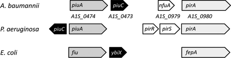
Chromosomal loci of piuA and pirA genes in A. baumannii, P. aeruginosa, and E. coli. The TBDR gene piuA is colocalized with the Fe(II)-dependent oxidoreductase gene piuC, while the pirA loci differ among the three organisms. Only genes relevant to the study context are shown.
PiuA and PirA contribute similarly to siderophore–beta-lactam activity in A. baumannii.
To test the involvement of PiuA and PirA in the activity of BAL30072 and MC-1 against A. baumannii, we constructed piuA (A1S_0474) and pirA (A1S_0980) gene deletion mutants in A. baumannii strain ATCC 19606. Both the piuA and the pirA mutants showed 2- to 8-fold increases in the MICs for both drug conjugates, in agreement with decreased transport in the individual mutants. The piuA-pirA double mutant showed a further increase in MICs, which was more pronounced for MC-1 than for BAL30072 (Table 2). This suggests that in the wild-type strain and under the conditions of the MIC assay, both receptors have a basal expression level and may contribute similarly to the uptake of BAL30072 and MC-1. This is in contrast to the situation in P. aeruginosa, since deletion of pirA in PAO1 has no effect on susceptibility to BAL30072 and MC-1, and the contribution of this TBDR is seen only in a piuA deletion background (15).
TABLE 2.
Susceptibilities of piuA and pirA deletion mutants of A. baumannii
| Strain | MIC (mg/liter) |
||
|---|---|---|---|
| BAL30072 | MC-1 | ATM | |
| ATCC 19606 | 0.25 | 1 | 32 |
| ATCC 19606 ΔpiuA | 1 | 2 | 32 |
| ATCC 19606 ΔpirA | 1–2 | 4 | 32 |
| ATCC 19606 ΔpirA ΔpiuA | 2–4 | 32 | 32 |
PiuA and PirA from A. baumannii are functional in P. aeruginosa.
To test whether the A. baumannii proteins are functional in P. aeruginosa, plasmids ppirA-Ab4.1 and ppiuA-Ab6.3, expressing, respectively, the pirA and piuA genes of A. baumannii from a constitutive or an inducible promoter, were transferred into P. aeruginosa strain PAO1 and into a piuA deletion mutant. While expression of the vector plasmids alone had no significant effect on the BAL30072 or MC-1 MIC, the expression of both piuA and pirA from A. baumannii conferred increased susceptibility to these conjugates in P. aeruginosa. As expected, susceptibility to aztreonam (ATM) was not affected by overexpression of the TBDR genes (Table 3). Similar results were obtained in a piuA deletion strain showing a 32- to 64-fold increase in susceptibility to BAL30072 and MC-1 when the pirA gene of either P. aeruginosa or A. baumannii was overexpressed. However, Ab-PiuA expression from plasmid ppiuA-Ab6.3 did not seem to confer increased susceptibility on the piuA deletion strain (Table 3). A possible explanation is that the induced expression level of Ab-PiuA in the piuA deletion strain might be insufficient to restore wild-type susceptibility.
TABLE 3.
Effects of heterologous expression of Ab-PiuA and Ab-PirA in P. aeruginosa PAO1 and PAO1 ΔpiuA on susceptibility to monocyclic beta-lactam antibiotics
| Strain/plasmid | MIC (mg/liter)a |
||
|---|---|---|---|
| BAL30072 | MC-1 | ATM | |
| PAO1 | 1–2 | 0.5 | 4 |
| pEX1.8 | 2 | 0.25 | 4 |
| pIApX2 | 1 | 0.5 | 8 |
| ppiuA1.1 (P. aeruginosa) | 0.125 | ND | 4 |
| ppiuAΔL1.1 (P. aeruginosa) | 2 | ND | 4 |
| ppiuA-Ab6.3 | 0.25 | 0.06 | 4 |
| ppirA1.1 (P. aeruginosa) | 0.25 | ND | 8 |
| ppirA-Ab4.1 | 0.06 | 0.03 | 4 |
| PAO1 ΔpiuA | 8–16 | 2 | 4 |
| pIApX2 | 16 | 4 | 8 |
| pEX1.8 | 16 | 4 | 4 |
| ppiuA1.1 (P. aeruginosa) | 0.125 | 0.03 | 8 |
| ppiuA-Ab6.3 | 8 | 2 | 2 |
| ppirA1.1 (P. aeruginosa) | 0.25 | 0.06 | 8 |
| ppirA-Ab4.1 | 0.25 | 0.125 | 4 |
Determined in MH medium supplemented with carbenicillin at 100 mg/liter and with 1 mM IPTG for strains harboring pEX1.8 and ppiuA-Ab6.3. ND, not done.
Structure comparison of PiuA and PirA from P. aeruginosa and A. baumannii.
The sequence alignment of the PiuA and PirA orthologues revealed high conservation within the putative substrate binding domains NL1 (PiuA and PirA) and NL2 and NL3 (PirA) (Fig. 2). The structures of PiuA and PirA from P. aeruginosa and A. baumannii have been solved. As expected, these structures were similar to those of other TBDRs, sharing a two-domain organization consisting of a 22-stranded transmembrane β-barrel (Fig. 3, green ribbons) and an N-terminal plug domain (blue ribbons) folded inside the barrel.
FIG 2.

Structure-based sequence alignment of PiuA and PirA proteins from P. aeruginosa and A. baumannii. Secondary-structure elements of Ab-PiuA are illustrated above the alignment. Regions predicted to be involved in the binding of the siderophores are boxed in red. Residues spatially equivalent to those involved in substrate binding in FhuA, FptA, and FpvA are highlighted in green. Key residues of the secondary binding site of FepA and conserved in the PirA structure are shown in yellow. Positions of residues of the nonconserved initial binding site are indicated by orange triangles. The plug domain is boxed in cyan. The alignment was drawn using Aline (71).
FIG 3.

Crystal structures of PiuA and PirA from P. aeruginosa and A. baumannii. β-Barrels are colored green, and N-terminal plug domains are blue. Loops NL1, NL2, and NL3 of the plug domains are shown in red. Residues likely to be part of the siderophore substrate binding site are shown as yellow sticks. The last solved residues of the flexible loops are represented as spheres.
As often observed with TBDRs, several extracellular loops were missing in the Pa-PiuA structure. Loops NL1 (83 to 90), L7 (505 to 526), and L8 (560 to 567) could not be modeled due to the high flexibility of these regions. In contrast, the equivalent extracellular loops were present in the structure of Ab-PiuA, perhaps due to the presence of a phosphate ion that coordinated with Glu73 of NL1, Asp303 of L4, Arg316 of β8, and His697 of L11. Structural comparison of Pa-PiuA and Ab-PiuA (root mean square deviations [RMSD] of 1.3 Å for 616 aligned C-α atoms) showed differences in the lengths and positions of several extracellular loops (Fig. 4; see Fig. S1 in the supplemental material). Ab-PiuA contained additional amino acids in loops L7 and L9, but fewer in L6 and L8 than Pa-PiuA. Strikingly, loop L7 of Ab-PiuA folded inside the barrel close to the top of the plug domain (Fig. 4; see Fig. S1 in the supplemental material). A mutant protein with five amino acids (EGGNP; positions 73 to 76) deleted from loop NL1 was generated in PiuA from P. aeruginosa. When expressed in strain PAO1, the NL1 deletion mutant was unable to confer hypersusceptibility to BAL30072, as observed with the PiuA wild-type protein (Table 3). We conclude that loop NL-1 is likely involved in the initial binding of BAL30072 in PiuA.
FIG 4.

Putative binding sites in PiuA of P. aeruginosa and A. baumannii and PirA of A. baumannii. The color scheme is the same as that in Fig. 3. The phosphate ion present in the Ab-PiuA structure is colored orange and red, and residues Asp303 and Glu73, which coordinate it, are represented as green and white sticks.
Comparison with other TBDRs showed that the closest structural relatives of PiuA are the pyochelin (FptA) and pyoverdine (FpvA) receptors of P. aeruginosa (37, 38), with an RMSD of 1.84 Å (567 aligned C-α atoms) and 1.99 Å (555 aligned C-α atoms), respectively, and the E. coli ferrichrome iron receptor FhuA, with an RMSD of 1.83 Å (600 aligned C-α atoms) (39, 40) (see Fig. S1 in the supplemental material).
As for PiuA, the extracellular loops of PirA from P. aeruginosa were highly flexible, and the structural analysis of Pa-PirA was focused on the structure of PirA from A. baumannii, where these loops where experimentally located. PirA was similar to the ferric enterobactin receptor FepA of E. coli (41), with an RMSD of 1.09 Å (618 aligned C-α atoms), as expected from the elevated sequence similarity (51% amino acid identity) (see Fig. S1 in the supplemental material). This is consistent with a previous report identifying PirA of P. aeruginosa as a secondary transporter of ferric enterobactin (36).
Selection of A. baumannii mutants with decreased susceptibility to BAL30072.
We investigated the possibility of resistance emergence in A. baumannii. Colonies of strains 307-0294 and ATCC 19606 able to grow in the presence of BAL30072 concentrations of 4 or 8 μg/ml were obtained at frequencies of 7 × 10⁻⁸ to 7 × 10⁻⁹. After determining their susceptibility profiles, we selected colonies that presented increased MICs of BAL30072 and MC-1 but unchanged MICs of ATM. Mutations in such colonies are expected to affect mechanisms specific to the siderophore-drug conjugate. Three out of 6 clones (R4, R5, and R6) obtained with strain 307-0294 and none of the 8 clones tested from strain ATCC 19606 fulfilled this criterion (Table 4). Therefore, clones R4, R5, and R6 were selected for further analysis and submitted to whole-genome sequencing. Mapping of the reads on the genome of strain 307-0294 revealed a single but distinct mutation in each of the three strains. Mutant R4 had acquired a frameshift mutation (insertion of A at position 9) in the exbD3 gene, while mutations in clones R5 (deletion of A at position 319) and R6 (insertion of A at position 243) resulted in frameshift mutations in the tonB3 gene (Table 4). Interestingly, the tonB3 gene, but not the tonB1 and tonB2 genes (see Fig. S2 in the supplemental material), has recently been shown to be induced under iron-limiting conditions (42). The TonB3-ExbB3-ExbD3 system could therefore be a likely candidate for providing energy to PiuA and PirA siderophore receptors in A. baumannii.
TABLE 4.
Susceptibilities of A. baumannii mutants selected on BAL30072
| Strain | MIC (mg/liter) |
Mutationa | ||
|---|---|---|---|---|
| BAL30072 | MC-1 | ATM | ||
| 307-0294 | 0.25 | 1 | 32 | WT |
| 307-0294-R4 | >8 | >8 | 16 | ExbD3 (Q4fs) |
| 307-0294-R5 | >8 | >8 | 16 | TonB3 (K106fs) |
| 307-0294-R6 | >8 | >8 | 16 | TonB3 (K80fs) |
fs, frameshift; WT, wild type.
Decreased growth rates of spontaneous exbD3 and tonB3 mutants of A. baumannii.
We observed that the spontaneous mutants R4, R5, and R6 formed slightly smaller colonies than the wild type when grown on lysogeny broth (LB) agar plates. To assess whether the mutations in the energy-transducing TonB3 system impose a fitness cost, we compared the growth of the mutants to that of the wild-type strain in Mueller-Hinton (MH) broth. As shown in Fig. 5, the tonB3 (R5 and R6) and exbD3 (R4) mutants were characterized by a longer lag phase and a lower final cell density after 20 h growth than the wild type. When the growth medium was supplemented with Fe(II)Cl2 at a final concentration of 20 μM, the growth phenotype of mutants R4 to R6 could be restored to reach nearly wild-type levels. The data suggest that TonB3-ExbB3-ExbD3 is important for siderophore-mediated Fe3+ acquisition and that the fitness of such mutants is likely to be severely decreased in vivo, where iron availability is limited.
FIG 5.
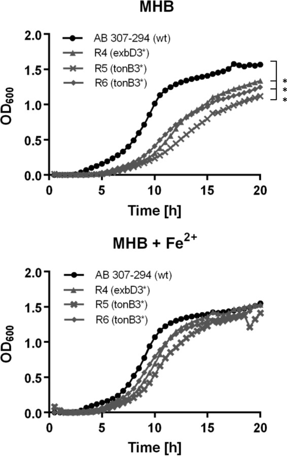
Growth of spontaneous mutants with decreased susceptibility to BAL30072. The strains were grown in MH broth (MHB) with or without Fe(II)Cl2 supplementation (20 μM final concentration). Growth was followed in a plate reader with regular OD600 readings. The data represent the average OD600 value for duplicate cultures. Experiments were repeated on three different occasions, yielding similar results. Data from one representative experiment are shown. The OD600 values, measured after 18 h for each of the three mutants, were statistically different from that of the wild type (Student t test; P < 0.05) in the absence, but not in the presence, of iron, as indicated by asterisks (top).
DISCUSSION
In this study, we have identified two siderophore receptors, termed Ab-PiuA and Ab-PirA, that are very likely involved in transport of the siderophore-monosulfactam conjugates BAL30072 and MC-1 into A. baumannii. The numbers of TBDRs found in different strains of A. baumannii range from 21 to 27 (compared to 35 in P. aeruginosa) (26). The close phylogenetic relationship between Acinetobacter and Pseudomonas prompted us to search in A. baumannii for orthologues of the previously identified TonB-dependent receptors PiuA and PirA from P. aeruginosa (15, 31). For each of these proteins, we identified one orthologue (>40% amino acid identity). The highest similarity was found within the putative substrate binding sites located in the N-terminal regions of the TBDRs (Fig. 2).
A transcriptome analysis performed in A. baumannii strain ATCC 17978 recently showed that expression of both piuA and pirA is induced under iron-limiting conditions (33). A. baumannii strains harbor at least three distinct siderophore clusters, one of them being the acinetobactin biosynthesis operon. However, neither PiuA nor PirA is part of these clusters. This suggests that the two TBDRs are likely involved in the uptake of xenosiderophores, although the substrate(s) of PiuA has not been identified. The closest orthologue of PiuA in E. coli is Fiu (35% amino acid identity), a TBDR also involved in the uptake of cephalosporins conjugated to catechol siderophores (12, 13).
PirA is an orthologue of the enterobactin receptor FepA in E. coli (51% amino acid identity). Enterobactin is a tricatechol siderophore produced by several enterobacterial species. However, the FepA receptor does not seem to play a role in the transport of catechol-based siderophore-drug conjugates in E. coli (12, 13), but it can transport beta-lactams linked to enterobactin (43, 44).
It is assumed in the design philosophy that BAL30072 interacts with the siderophore receptors PiuA and PirA in a manner similar to that of their natural siderophores. However, the structures of the loops differ between the orthologous proteins from the two organisms. There are even larger differences between the loop structures of PirA and PiuA (see Fig. S1 in the supplemental material). Since the natural siderophores of PiuA and PirA are not known, we rely on comparisons with known complexes to inform us as to the recognition elements. The binding site of known siderophore receptors is in general formed by loops NL1, NL2, and NL3 of the plug domain and loops L3, L7, and L11 and strands β7, β8, and β9 of the β-barrel (Fig. 2) (37, 39, 45).
The closest structural homologues of PiuA are FptA, FpvA, and FhuA, and their complexes with cognate siderophores show a cluster of aromatic and positively charged residues (Fig. 4). The side chains of Tyr239 on loop L3 of Pa-PiuA and Trp298 in β7 of Ab-PiuA have positions similar to those of Trp362 in FpvA and Met271 in FptA, both involved in siderophore binding. Tyrosines 307 and 296 on loop β7 of Pa-PiuA and Ab-PiuA occupy positions equivalent to those of FpvA Trp391 and FhuA Tyr116. A positively charged residue on β8 (Lys329 for Pa-PiuA and Arg316 for Ab-PiuA) is in the same position as the side chain of Lys344 in FhuA. Trp498, present in loop L7 of Ab-PiuA, occupies the same volume as Tyr231 and Phe115 in NL3 of FpvA and FhuA, respectively. A tyrosine-histidine couple (residues 694 and 697 in Ab-PiuA and 697 and 700 in Pa-PiuA) is superimposed with Phe795 and Tyr796 in FpvA and Phe704 and Tyr707 in FhuA. We note that a phosphate ion is found in the Ab-PiuA structure, where it is coordinated by Arg316 and His697. There are important differences, however, between Pa-PiuA and Ab-PiuA. In particular, loops NL2 and NL3 are shorter in Pa-PiuA, while NL1 extends above the plug domain of Ab-PiuA (Fig. 3). Consequently, NL1 in Ab-PiuA would clash with the siderophore location seen in FpvA, FptA, and FhuA (see Fig. S1 in the supplemental material). This may reflect a genuine difference with a functional consequence or simply an inactive conformation.
Although there is no structure for the siderophore complex of FepA from E. coli, several binding and mutation studies have been reported (46–48). These studies suggest a biphasic binding kinetic with an initial binding site in the loop extremities and a secondary site deeper inside the barrel leading to the translocation. Loops L2, L4, L7, L8, L10, and L11 were found to be involved in the binding of ferric enterobactin (41, 49). These loops are different in structure and sequence from those found in PirA, consistent with the observation that FepA does not transport BAL30072. Interestingly, it also indicates that PirA, which can recognize enterobactin, may use a different constellation of residues than FepA. The presumed second binding site, on top of the plug domain, is highly conserved between the two PirAs and FepA. Deletion of loop NL1 or NL3 in FepA or mutation of the conserved Trp101 in NL3 (corresponding to Trp111 and Trp110 in Ab-PirA and Pa-PirA) drastically decreased the uptake of ferric enterobactin (48, 49). Other key residues known to be important in FepA are Tyr260 of L3 (262 in Ab-PirA and 264 in Pa-PirA), Arg316 on β7 (Ab-PirA R320 and Pa-PirA R321), and Tyr478 of L7 (Ab-PirA Tyr478 and Pa-PirA Tyr479), which are conserved in PirA. As in FepA, loops L3, L4, L7, and L10 cover the top of the barrel, making the plug domain less accessible than for the other TBDRs.
TBDRs interact with the cytoplasmic-membrane protein TonB, which transmits energy derived from the proton gradient to the receptor via interaction with the N-terminal TonB box, to drive substrate transport (see Fig. S2 in the supplemental material). A. baumannii harbors three distinct chromosomally encoded TonB systems, among which TonB3-ExbB3-ExbD3 was recently shown to be iron regulated (42). Spontaneous resistant clones able to grow on >4 mg/liter of BAL30072 carried frameshift mutations in tonB3 or exbD3 (see Fig. S2 in the supplemental material). It is therefore highly likely that the TonB3-ExbB3-ExbD3 system interacts with PiuA and PirA to provide the energy for the uptake of BAL30072 and MC-1. Of note, the MICs of BAL30072 for the tonB3 and exbD3 mutants were higher than those for the individual TBDR piuA and pirA mutants or the pirA-piuA double mutant (compare the values in Tables 2 and 4). Although the strain backgrounds are different (ATCC 19606 versus 307-0294), it suggests that additional TBDRs could be involved in transport of siderophore-drug conjugates in A. baumannii strain 307-0294. This assumption would also explain the selection of exbD3 and tonB3 mutants at 4 mg/liter of BAL30072, which display higher MICs than the individual pirA and piuA mutants in strain ATCC 19606.
The A. baumannii tonB3 and exbD3 mutants selected in strain 307-0294 on BAL30072 showed decreased growth rates under iron-limiting conditions. A similar growth impairment under iron deficiency was reported by Zimbler et al. for tonB1 and tonB2 deletion mutants of strain ATCC 19606 (42). Since the authors of the study could not generate a tonB3 deletion mutant by homologous recombination, the growth phenotype could not be assessed, and the authors concluded the TonB3-ExbB3-ExbD3 system was essential in A. baumannii. Whether this was due to the experimental conditions or to differences in strain backgrounds is unclear. Supplementation with iron could restore the growth defects in their tonB1 and tonB2 mutants (42), as well as in our spontaneous tonB3 and exbD3 mutants of strain 307-0294 (Fig. 5). It remains to be determined if tonB1 and tonB2 mutants also show altered susceptibilities to siderophore-drug conjugates. Altogether, the data suggest that tonB mutants, if selected in vivo, would be impaired in their fitness and probably also be less virulent in the patient, where iron availability is limited.
Both endogenous and heterologous overexpression of Ab-PiuA and Ab-PirA conferred increased susceptibility to both BAL30072 and MC-1 on P. aeruginosa strain PAO1 (Table 3). This establishes the ability of these two proteins to transport the two antibiotics; moreover, it indicates that the amounts of PiuA and PirA expressed in PAO1 are probably the bottleneck for the transport of siderophore-drug conjugates, rather than the availability of the TonB-ExbB/ExbD systems. There must be mutual recognition between the C-terminal part of the cytoplasmic-membrane protein TonB in P. aeruginosa and the TonB box located in the N-terminal domains of PirA and PiuA from A. baumannii. In P. aeruginosa, the energy coupling to iron-transporting TBDRs is mediated by TonB1 (50), while our data suggest that TonB3 is the equivalent protein in A. baumannii. However, TonB1 (P. aeruginosa) and TonB3 (A. baumannii) show only limited amino acid sequence identity. The TonB3 domain of A. baumannii displayed the highest sequence similarity to the C-terminal regions of TolA (amino acids [aa] 277 to 322; 48% identity) and TonB2 (aa 178 to 223; 39% identity) of P. aeruginosa. These domains are located in the periplasm and are thought to interact with the corresponding TBDR in the outer membrane (51). Which TonB protein of P. aeruginosa is interacting with PiuA and PirA from A. baumannii remains to be determined. To our knowledge, this is the first report on heterologous complementation between TonB-dependent receptors in Gram-negative nonfermenters.
In summary, the previously uncharacterized TBDRs PiuA and PirA are the major uptake systems for the siderophore-drug conjugates BAL30072 and MC-1 in A. baumannii. Despite their overall similar crystal structures, differences in the external loops of the plug domain could be important for substrate selectivity. We obtained evidence for heterologous recognition between a TonB protein(s) of P. aeruginosa and TBDRs of A. baumannii. Our study highlights the potential of siderophore-drug conjugates as an efficient strategy to increase antimicrobial transport into Gram-negative bacteria.
MATERIALS AND METHODS
Bacterial strains and growth conditions.
The strains and plasmids used in this study are listed in Table 1, and the primers used are shown in Table S1 in the supplemental material. A. baumannii strains were grown in double-strength yeast tryptone (dYT) broth and E. coli and P. aeruginosa in LB at 37°C with shaking (220 rpm). E. coli XL-1 Blue and DH10B were used as cloning hosts. E. coli ST18 was the donor strain for biparental matings, requiring supplementation with 50 μg/ml 5-aminolevulinic acid. Gentamicin (25 μg/ml) or kanamycin (25 μg/ml) was added for plasmid-carrying strains. MIC determinations were performed in MH broth according to CLSI guidelines (52) and were repeated on at least three different occasions.
Mutant selection and growth assay.
Spontaneous mutants of the fully sequenced A. baumannii strain 307-0294 were selected on LB agar plates supplemented with BAL30072. Individual colonies were picked and grown in the absence of antibiotic. MICs were determined, and strains with decreased susceptibility to BAL30072 were retained for further analysis. The growth of mutant strains was performed in microtiter plates in a total volume of 200 μl. Strains were grown for 7 h in LB with shaking (250 rpm) at 37°C. Cultures were diluted to an optical density at 600 nm (OD600) of 0.015, and 4 μl was inoculated into 196 μl of MH broth. Growth was followed in a BioTek Synergy 1 plate reader at 37°C with shaking for 1 min before hourly measurement of the optical density at 600 nm. The growth curves represent the average OD600 values for duplicate cultures.
PCR amplifications and DNA modifications.
PCR primers are listed in Table S1 in the supplemental material. All primer sequences were based on the genome of A. baumannii ATCC 19606. Screening PCRs were carried out using the DreamTaq DNA polymerase (Thermo Scientific), in accordance with the manufacturer's instructions and the optimized annealing temperature for each primer set. For screening PCRs, bacterial cells were boiled at 95°C for 5 min and subsequently pelleted at 13,000 rpm for 1 min. Phusion DNA polymerase (Thermo Scientific) was used for high-fidelity PCRs (supplemented with 5% dimethyl sulfoxide [DMSO]). Restriction digestions were performed using Thermo Scientific restriction enzymes, according to the manufacturer's instructions, at the appropriate temperature. All ligation reactions were carried out at room temperature using Thermo Scientific T4 DNA ligase. DNA purifications were performed using either the GeneJet PCR purification or the GeneJet gel extraction kit (Thermo Scientific), following the manufacturer's instructions.
Construction of pirA and piuA knockout mutants in A. baumannii.
The generation of unmarked knockout mutants was based on the protocol described by Hoang et al. (53). Briefly, DNA fragments of 500 to 700 bp were PCR amplified using primer pairs A1/A2 and B1/B2, respectively (see Table S1 in the supplemental material). For deletion of pirA, the up- and downstream regions flanking the gene were PCR amplified. For knockout of piuA, the amplified fragments were located in the 5′ and 3′ regions of the genes. After amplification, the obtained A and B fragments were gel purified, and approximately 40 ng of each fragment was used in a PCR fusion with primers A1 and B2, which share an 18-bp homologous region. The resulting fusion products were gel purified and further cloned into the suicide vectors pEX18Gm, via HindIII/EcoRI restriction sites (pirA), and pEX18Km, via SalI/EcoRI (piuA). The cloned knockout fragments were verified by sequencing. The replacement vectors were mobilized into A. baumannii via biparental conjugation, and the generation of the unmarked mutants was carried out as previously described (54). In order to confirm a gene deletion, locus-specific primers (out1 and out2) that bind up- and downstream of the gene were designed and used for amplification of the knockout region. Sequencing of the obtained PCR fragments confirmed successful deletion of the genes.
Construction of expression plasmids.
The piuA and pirA coding regions were amplified by PCR from genomic DNA of P. aeruginosa reference strain PAO1. The piuA coding region was amplified with primers piuA-Xba and piuA-Hind and cloned as a 2,540-bp XbaI-HindIII DNA fragment into the expression vector pIApX2, yielding plasmid ppiuA1.1. Deletions of residues 73 to 76 from loop NL1 (ppiuAΔL1.1) were constructed with a modified protocol of the QuikChange method (55) using primers piuA-Δ1 and piuA-Δ2. The pirA coding region was amplified as a 2,498-bp DNA fragment obtained with primers pirA-Bam and pirA-Hind2 (see Table S1 in the supplemental material). The PCR product was digested by BamHI and HindIII and cloned into pIApX2, yielding plasmid ppirA1.1. The coding regions of piuA (A1S_0474) and pirA (A1S_0980) were amplified by PCR from genomic DNA of A. baumannii strain 307-0294 (provided by T. A. Russo, SUNY, Buffalo, NY, USA), using primers listed in Table S1 in the supplemental material. The 2,436-bp-long piuA fragment was digested with EcoRI and PstI and ligated to the EcoRI- and PstI-cleaved vector pEX1.8, yielding plasmid ppiuA-Ab6.3. The 2,557-bp-long pirA fragment was cleaved with XbaI and PstI and ligated to plasmid pIApX2 cleaved with the same restriction enzymes, yielding plasmid ppirA-Ab4.1. The plasmids were transferred into P. aeruginosa by electroporation. The piuA gene of A. baumannii (piuA-Ab) was further subcloned by BamHI-PstI digestion into the A. baumannii expression vector pBAV1K-5T-gfp (56) (kindly provided by G. Wilharm, Robert Koch Institut, Werningerode, Germany), yielding plasmid ppiuA-Ab7.1. The piuA-Ab gene was further subcloned by XbaI-PstI digestion into pBAV1K-5T-gfp, yielding plasmid ppirA-Ab3.1. The PCR conditions were as follows: denaturation at 95°C for 2 min, followed by 27 cycles of 95°C for 20 s, 57°C for 30 s, and 72°C for 2 min and a final extension at 72°C for 4 min. Plasmids were introduced into P. aeruginosa and A. baumannii by electroporation. Plasmid-containing colonies were selected on LB agar supplemented with carbenicillin at 200 mg/liter (P. aeruginosa) or kanamycin at 50 mg/liter (A. baumannii).
Cloning, overexpression, and purification of PiuA and PirA from P. aeruginosa and A. baumannii.
The positions of the signal peptide cleavage sites of the proteins were predicted with Signal P4.0 (57). The coding sequence of the mature protein Pa-PiuA (PA4514) was amplified from PAO1 genomic DNA using primer pairs Pa-PiuA-F/Pa-PiuA-R (see Table S1 in the supplemental material) and Pfu DNA polymerase. The PCR product was digested by BspHI and EagI restriction enzymes and cloned into vector pET20b using restriction enzymes NcoI and EagI so that the protein was expressed with a PelB signal sequence for the outer membrane localization and a noncleavable C-terminal His6 tag. Codon-optimized Pa-PirA (PA0931) with a C-terminal, noncleavable His6 tag was purchased from DNA2.0 and inserted into vector pET20b using HindIII and EcoRV restriction sites.
The coding sequences of the mature proteins Ab-PiuA and Ab-PirA were amplified by PCR from A. baumannii strain ATCC 19606 using KOD DNA polymerase (Novagen) and the primer pairs Ab-PiuA-F/Ab-PiuA-R and Ab-PirA-F/Ab-PirA-R (see Table S1 in the supplemental material). Both genes were cloned with an N-terminal tobacco etch virus (TEV) protease-cleavable His7 tag in vector pTAMAHISTEV using NcoI and BamHI restriction sites. The pTAMAHISTEV expression vector was obtained by replacing the PelB signal peptide of plasmid pEPELBHISTEV (courtesy of Huanting Liu, University of St Andrews, St Andrews, United Kingdom) with the TamA signal peptide, as described by Liu et al. (58).
Proteins were overexpressed in E. coli strain C43(DE3) or BL21(DE3). Cells were grown at 37°C in LB medium containing 100 mg/liter ampicillin to an OD600 of 0.7 and then induced with 0.4 mM isopropyl-β-d-thiogalactopyranoside (IPTG) at 25°C overnight.
For Pa-PiuA and Pa-PirA, harvested cells were resuspended in lysis buffer (50 mM sodium phosphate, pH 7.5, 250 mM NaCl, 10% glycerol, 100 μg/ml lysozyme, and 20 μg/ml DNase). The cells were lysed by two passages through a cell disruptor at 30,000 lb/in2 (Constant Systems). Unbroken cells were removed by centrifugation (20 min at 10,000 × g), and membrane fractions were isolated by ultracentrifugation (100,000 × g; 1 h). The supernatant was discarded, and inner membrane proteins were solubilized in buffer 1 (50 mM sodium phosphate, pH 7.5, 250 mM NaCl, 10% glycerol, and 1% [wt/vol] N-lauroylsarkosine [Sigma]). The insoluble outer membrane fraction was isolated by ultracentrifugation (100,000 × g; 1 h), and outer membrane proteins were solubilized in buffer 2 (50 mM sodium phosphate, pH 8, 250 mM NaCl, and 7% [vol/vol] octylpolyoxyethylene [octylPOE] [Bachem]). Insoluble material was removed by ultracentrifugation (100,000 × g; 1 h). The supernatant was applied to a charged Histrap nickel-sepharose high-performance column (GE Healthcare) equilibrated with 1% (vol/vol) octylPOE, 250 mM NaCl, 10 mM imidazole, and 50 mM sodium phosphate, pH 8, and washed with the same buffer containing 15 mM imidazole. Protein was eluted with a linear gradient of imidazole (5 to 150 mM). Fractions containing the protein were pooled; dialyzed against 50 mM Tris, pH 8.4, and 1% octylPOE; loaded on an anion exchange column (Resource Q [GE Healthcare]) equilibrated with the dialysis buffer; and eluted with a linear gradient of 0 to 600 mM NaCl over 12 column volumes. Fractions were pooled and loaded on a Superdex S200 gel filtration column (GE Healthcare) equilibrated with 10 mM Tris, pH 8, 150 mM NaCl, and 0.45% (vol/vol) n-octyltetraoxyethylene (C8E4) (Bachem). Protein fractions were pooled and concentrated to 8 mg/ml.
The expression and initial purification steps for Ab-PiuA and Ab-PirA were the same as for Pa-PiuA and Pa-PirA. After applying them to a first immobilized-metal affinity chromatography (IMAC) column, imidazole was removed by dialysis (50 mM Tris, pH 8, 250 mM NaCl, 10% glycerol, 2 mM mercaptoethanol, and 1% octylPOE) before adding TEV protease to cleave the His7 tag. The protein was applied to a second IMAC column, followed by Superdex S200 gel filtration chromatography (GE Healthcare) (10 mM Tris, pH 8, 150 mM NaCl, 0.45% C8E4). The protein fractions were pooled and concentrated to 10 mg/ml.
Crystallization and structure determination.
Crystals of Pa-PiuA appeared at 20°C after several days by mixing 1 μl of protein solution (8 mg/ml) with 1 μl of reservoir solution containing 32% polyethylene glycol (PEG) 400, 0.05 M Tris, pH 8.5, 0.05 M sodium sulfate, and 0.05 M lithium chloride. Pa-PirA crystals were obtained at 20°C by mixing 1.5 μl of the protein solution (7.5 mg/ml) containing 2.5 mM n-dodecylphosphocholine (Anatrace) with 1 μl of reservoir solution containing 17% PEG 3350, 0.2 M magnesium chloride, 0.1 M Tris, pH 7. The crystals were frozen with the same solution containing 25% PEG 400.
Crystals of Ab-PiuA appeared at 20°C after several days by mixing 1 μl of protein solution (10 mg/ml) with 1 μl of reservoir solution containing 1 M diammonium hydrogen phosphate, 0.1 M sodium acetate, pH 4.6. The crystals were cryoprotected by supplementing the mother liquor with 30% sucrose. Prior to crystallization, Fe3+-enterobactin was added to Ab-PirA at a final concentration of 1 mM before loading on a Superdex S200. Ab-PirA crystals appeared a week after mixing at 20°C 1 μl of protein solution (7 mg/ml) with 1 μl of reservoir solution containing 16% PEG 8000, 0.15 M magnesium sulfate, and 0.1 M bicine, pH 9.
Data were collected at beamlines IO4 and IO3 at the Diamond Light Source, Oxfordshire, United Kingdom, and ID14-4 at European Synchrotron Radiation Facility (ESRF). Data were processed with XIA2 (59–63). The structure of Pa-PiuA was solved by molecular replacement using the coordinates of FhuA (Protein Data Bank [PDB] code 2FCP) (39) as a model with the program PHASER (64). The structure of Ab-PiuA was solved using Pa-PiuA coordinates, and the structures of Pa-PirA and Ab-PirA were solved using FepA coordinates (PDB code 1FEP) (41). Models were adjusted with COOT (65), and refinement was carried out using REFMAC in the CCP4 program suite with TLS parameters (66). The final refinement statistics are given in Table S2 in the supplemental material. The quality of all structures was checked with MOLPROBITY (67). Figures were drawn using PyMOL (version 1.8; Schrödinger, LLC).
Whole-genome sequencing.
Samples were sequenced on an Illumina MiSeq V2 instrument. Assembly and analysis were performed off instrument using CLCBio Genomics Workbench v 6.5 (Cambridge, MA). Detection of single nucleotide polymorphisms (SNPs)/indels was accomplished through mapping to a parent reference assembly using the same parameters. Quality-based SNPs were detected at a minimum frequency of 80% using default criteria and verified by examination of de novo assembly. Annotation of parental strains was accomplished through ORF calling using Prodigal software and BLAST analysis at NCBI for regions of interest. Sequence analyses were performed using the BLAST algorithm on genomic data provided by the MIST2.2 database (mistdb.com).
Accession number(s).
Atomic coordinates and structure factors have been deposited in the Protein Data Bank (accession no. 5FOK, 5FP2, 5FP1, and 5FR8).
Supplementary Material
ACKNOWLEDGMENTS
We are grateful to R. A. Alm and S. Mills from Astra Zeneca (Waltham, MA, USA), who performed the whole-genome sequencing of the A. baumannii mutants.
The research leading to these results was conducted as part of the Translocation consortium and has received support from the Innovative Medicines Joint Undertaking under grant agreement 115525, resources which are composed of financial contribution from the European Union's seventh framework program (FP7/2007-2013) and EFPIA companies in kind contribution.
Footnotes
Supplemental material for this article may be found at https://doi.org/10.1128/AAC.02531-16.
REFERENCES
- 1.Li XZ, Plesiat P, Nikaido H. 2015. The challenge of efflux-mediated antibiotic resistance in Gram-negative bacteria. Clin Microbiol Rev 28:337–418. doi: 10.1128/CMR.00117-14. [DOI] [PMC free article] [PubMed] [Google Scholar]
- 2.Ji C, Juarez-Hernandez RE, Miller MJ. 2012. Exploiting bacterial iron acquisition: siderophore conjugates. Future Med Chem 4:297–313. doi: 10.4155/fmc.11.191. [DOI] [PMC free article] [PubMed] [Google Scholar]
- 3.Schalk IJ, Mislin GL, Brillet K. 2012. Structure, function and binding selectivity and stereoselectivity of siderophore-iron outer membrane transporters. Curr Top Membr 69:37–66. doi: 10.1016/B978-0-12-394390-3.00002-1. [DOI] [PubMed] [Google Scholar]
- 4.Schauer K, Rodionov DA, de Reuse H. 2008. New substrates for TonB-dependent transport: do we only see the ‘tip of the iceberg’? Trends Biochem Sci 33:330–338. doi: 10.1016/j.tibs.2008.04.012. [DOI] [PubMed] [Google Scholar]
- 5.Neidhardt FC, Bloch PL, Smith DF. 1974. Culture medium for enterobacteria. J Bacteriol 119:736–747. [DOI] [PMC free article] [PubMed] [Google Scholar]
- 6.Reynolds PR, Mottur GP, Bradbeer C. 1980. Transport of vitamin B12 in Escherichia coli. Some observations on the roles of the gene products of BtuC and TonB. J Biol Chem 255:4313–4319. [PubMed] [Google Scholar]
- 7.Wencewicz TA, Miller MJ. 2013. Biscatecholate-monohydroxamate mixed ligand siderophore-carbacephalosporin conjugates are selective sideromycin antibiotics that target Acinetobacter baumannii. J Med Chem 56:4044–4052. doi: 10.1021/jm400265k. [DOI] [PMC free article] [PubMed] [Google Scholar]
- 8.Ji C, Miller PA, Miller MJ. 2012. Iron transport-mediated drug delivery: practical syntheses and in vitro antibacterial studies of tris-catecholate siderophore-aminopenicillin conjugates reveals selectively potent antipseudomonal activity. J Am Chem Soc 134:9898–9901. doi: 10.1021/ja303446w. [DOI] [PMC free article] [PubMed] [Google Scholar]
- 9.Maejima T, Inoue M, Mitsuhashi S. 1991. In vitro antibacterial activity of KP-736, a new cephem antibiotic. Antimicrob Agents Chemother 35:104–110. doi: 10.1128/AAC.35.1.104. [DOI] [PMC free article] [PubMed] [Google Scholar]
- 10.Kohira N, West J, Ito A, Ito-Horiyama T, Nakamura R, Sato T, Rittenhouse S, Tsuji M, Yamano Y. 2016. In vitro antimicrobial activity of siderophore cephalosporin S-649266 against Enterobacteriaceae clinical isolates including carbapenem-resistant strains. Antimicrob Agents Chemother 60:729–734. doi: 10.1128/AAC.01695-15. [DOI] [PMC free article] [PubMed] [Google Scholar]
- 11.Ito A, Kohira N, Bouchillon SK, West J, Rittenhouse S, Sader HS, Rhomberg PR, Jones RN, Yoshizawa H, Nakamura R, Tsuji M, Yamano Y. 2016. In vitro antimicrobial activity of S-649266, a catechol-substituted siderophore cephalosporin, when tested against non-fermenting Gram-negative bacteria. J Antimicrob Chemother 71:670–677. doi: 10.1093/jac/dkv402. [DOI] [PubMed] [Google Scholar]
- 12.Nikaido H, Rosenberg EY. 1990. Cir and Fiu proteins in the outer membrane of Escherichia coli catalyze transport of monomeric catechols: study with beta-lactam antibiotics containing catechol and analogous groups. J Bacteriol 172:1361–1367. doi: 10.1128/jb.172.3.1361-1367.1990. [DOI] [PMC free article] [PubMed] [Google Scholar]
- 13.Curtis NA, Eisenstadt RL, East SJ, Cornford RJ, Walker LA, White AJ. 1988. Iron-regulated outer membrane proteins of Escherichia coli K-12 and mechanism of action of catechol-substituted cephalosporins. Antimicrob Agents Chemother 32:1879–1886. doi: 10.1128/AAC.32.12.1879. [DOI] [PMC free article] [PubMed] [Google Scholar]
- 14.Page MG, Dantier C, Desarbre E. 2010. In vitro properties of BAL30072, a novel siderophore sulfactam with activity against multiresistant gram-negative bacilli. Antimicrob Agents Chemother 54:2291–2302. doi: 10.1128/AAC.01525-09. [DOI] [PMC free article] [PubMed] [Google Scholar]
- 15.McPherson CJ, Aschenbrenner LM, Lacey BM, Fahnoe KC, Lemmon MM, Finegan SM, Tadakamalla B, O'Donnell JP, Mueller JP, Tomaras AP. 2012. Clinically-relevant Gram-negative resistance mechanisms have no effect on the efficacy of MC-1, a novel siderophore-conjugated monocarbam. Antimicrob Agents Chemother 56:6334–6342. doi: 10.1128/AAC.01345-12. [DOI] [PMC free article] [PubMed] [Google Scholar]
- 16.Han S, Caspers N, Zaniewski RP, Lacey BM, Tomaras AP, Feng X, Geoghegan KF, Shanmugasundaram V. 2011. Distinctive attributes of beta-lactam target proteins in Acinetobacter baumannii relevant to development of new antibiotics. J Am Chem Soc 133:20536–20545. doi: 10.1021/ja208835z. [DOI] [PubMed] [Google Scholar]
- 17.Mima T, Kvitko BH, Rholl DA, Page MG, Desarbre E, Schweizer HP. 2011. In vitro activity of BAL30072 against Burkholderia pseudomallei. Int J Antimicrob Agents 38:157–159. doi: 10.1016/j.ijantimicag.2011.03.019. [DOI] [PMC free article] [PubMed] [Google Scholar]
- 18.Russo TA, Page MG, Beanan JM, Olson R, Hujer AM, Hujer KM, Jacobs M, Bajaksouzian S, Endimiani A, Bonomo RA. 2011. In vivo and in vitro activity of the siderophore monosulfactam BAL30072 against Acinetobacter baumannii. J Antimicrob Chemother 66:867–873. doi: 10.1093/jac/dkr013. [DOI] [PMC free article] [PubMed] [Google Scholar]
- 19.Dijkshoorn L, Nemec A, Seifert H. 2007. An increasing threat in hospitals: multidrug-resistant Acinetobacter baumannii. Nat Rev Microbiol 5:939–951. doi: 10.1038/nrmicro1789. [DOI] [PubMed] [Google Scholar]
- 20.Bou G, Martinez-Beltran J. 2000. Cloning, nucleotide sequencing, and analysis of the gene encoding an AmpC beta-lactamase in Acinetobacter baumannii. Antimicrob Agents Chemother 44:428–432. doi: 10.1128/AAC.44.2.428-432.2000. [DOI] [PMC free article] [PubMed] [Google Scholar]
- 21.Bonnin RA, Nordmann P, Poirel L. 2013. Screening and deciphering antibiotic resistance in Acinetobacter baumannii: a state of the art. Expert Rev Anti Infect Ther 11:571–583. doi: 10.1586/eri.13.38. [DOI] [PubMed] [Google Scholar]
- 22.Potron A, Poirel L, Nordmann P. 2015. Emerging broad-spectrum resistance in Pseudomonas aeruginosa and Acinetobacter baumannii: mechanisms and epidemiology. Int J Antimicrob Agents 45:568–585. doi: 10.1016/j.ijantimicag.2015.03.001. [DOI] [PubMed] [Google Scholar]
- 23.Magnet S, Courvalin P, Lambert T. 2001. Resistance-nodulation-cell division-type efflux pump involved in aminoglycoside resistance in Acinetobacter baumannii strain BM4454. Antimicrob Agents Chemother 45:3375–3380. doi: 10.1128/AAC.45.12.3375-3380.2001. [DOI] [PMC free article] [PubMed] [Google Scholar]
- 24.Damier-Piolle L, Magnet S, Bremont S, Lambert T, Courvalin P. 2008. AdeIJK, a resistance-nodulation-cell division pump effluxing multiple antibiotics in Acinetobacter baumannii. Antimicrob Agents Chemother 52:557–562. doi: 10.1128/AAC.00732-07. [DOI] [PMC free article] [PubMed] [Google Scholar]
- 25.Coyne S, Rosenfeld N, Lambert T, Courvalin P, Perichon B. 2010. Overexpression of resistance-nodulation-cell division pump AdeFGH confers multidrug resistance in Acinetobacter baumannii. Antimicrob Agents Chemother 54:4389–4393. doi: 10.1128/AAC.00155-10. [DOI] [PMC free article] [PubMed] [Google Scholar]
- 26.Koebnik R. 2005. TonB-dependent trans-envelope signalling: the exception or the rule? Trends Microbiol 13:343–347. doi: 10.1016/j.tim.2005.06.005. [DOI] [PubMed] [Google Scholar]
- 27.Funahashi T, Tanabe T, Mihara K, Miyamoto K, Tsujibo H, Yamamoto S. 2012. Identification and characterization of an outer membrane receptor gene in Acinetobacter baumannii required for utilization of desferricoprogen, rhodotorulic acid, and desferrioxamine B as xenosiderophores. Biol Pharm Bull 35:753–760. doi: 10.1248/bpb.35.753. [DOI] [PubMed] [Google Scholar]
- 28.Mihara K, Tanabe T, Yamakawa Y, Funahashi T, Nakao H, Narimatsu S, Yamamoto S. 2004. Identification and transcriptional organization of a gene cluster involved in biosynthesis and transport of acinetobactin, a siderophore produced by Acinetobacter baumannii ATCC 19606T. Microbiology 150:2587–2597. doi: 10.1099/mic.0.27141-0. [DOI] [PubMed] [Google Scholar]
- 29.Yamamoto S, Okujo N, Sakakibara Y. 1994. Isolation and structure elucidation of acinetobactin, a novel siderophore from Acinetobacter baumannii. Arch Microbiol 162:249–254. [DOI] [PubMed] [Google Scholar]
- 30.Zimbler DL, Penwell WF, Gaddy JA, Menke SM, Tomaras AP, Connerly PL, Actis LA. 2009. Iron acquisition functions expressed by the human pathogen Acinetobacter baumannii. Biometals 22:23–32. doi: 10.1007/s10534-008-9202-3. [DOI] [PubMed] [Google Scholar]
- 31.Van Delden C, Page MG, Kohler T. 2013. Involvement of Fe uptake systems and AmpC beta-lactamase in susceptibility to the siderophore monosulfactam BAL30072 in Pseudomonas aeruginosa. Antimicrob Agents Chemother 57:2095–2102. doi: 10.1128/AAC.02474-12. [DOI] [PMC free article] [PubMed] [Google Scholar]
- 32.Higgins PG, Stefanik D, Page MG, Hackel M, Seifert H. 2012. In vitro activity of the siderophore monosulfactam BAL30072 against meropenem-non-susceptible Acinetobacter baumannii. J Antimicrob Chemother 67:1167–1169. doi: 10.1093/jac/dks009. [DOI] [PubMed] [Google Scholar]
- 33.Eijkelkamp BA, Hassan KA, Paulsen IT, Brown MH. 2011. Investigation of the human pathogen Acinetobacter baumannii under iron limiting conditions. BMC Genomics 12:126. doi: 10.1186/1471-2164-12-126. [DOI] [PMC free article] [PubMed] [Google Scholar]
- 34.Aravind L, Koonin EV. 2001. The DNA-repair protein AlkB, EGL-9, and leprecan define new families of 2-oxoglutarate- and iron-dependent dioxygenases. Genome Biol 2:RESEARCH0007. [DOI] [PMC free article] [PubMed] [Google Scholar]
- 35.Zimbler DL, Park TM, Arivett BA, Penwell WF, Greer SM, Woodruff TM, Tierney DL, Actis LA. 2012. Stress response and virulence functions of the Acinetobacter baumannii NfuA Fe-S scaffold protein. J Bacteriol 194:2884–2893. doi: 10.1128/JB.00213-12. [DOI] [PMC free article] [PubMed] [Google Scholar]
- 36.Ghysels B, Ochsner U, Mollman U, Heinisch L, Vasil M, Cornelis P, Matthijs S. 2005. The Pseudomonas aeruginosa pirA gene encodes a second receptor for ferrienterobactin and synthetic catecholate analogues. FEMS Microbiol Lett 246:167–174. doi: 10.1016/j.femsle.2005.04.010. [DOI] [PubMed] [Google Scholar]
- 37.Cobessi D, Celia H, Pattus F. 2005. Crystal structure at high resolution of ferric-pyochelin and its membrane receptor FptA from Pseudomonas aeruginosa. J Mol Biol 352:893–904. doi: 10.1016/j.jmb.2005.08.004. [DOI] [PubMed] [Google Scholar]
- 38.Cobessi D, Celia H, Folschweiller N, Schalk IJ, Abdallah MA, Pattus F. 2005. The crystal structure of the pyoverdine outer membrane receptor FpvA from Pseudomonas aeruginosa at 3.6 angstroms resolution. J Mol Biol 347:121–134. doi: 10.1016/j.jmb.2005.01.021. [DOI] [PubMed] [Google Scholar]
- 39.Ferguson AD, Hofmann E, Coulton JW, Diederichs K, Welte W. 1998. Siderophore-mediated iron transport: crystal structure of FhuA with bound lipopolysaccharide. Science 282:2215–2220. doi: 10.1126/science.282.5397.2215. [DOI] [PubMed] [Google Scholar]
- 40.Krissinel E, Henrick K. 2004. Secondary-structure matching (SSM), a new tool for fast protein structure alignment in three dimensions. Acta Crystallogr D Biol Crystallogr 60:2256–2268. doi: 10.1107/S0907444904026460. [DOI] [PubMed] [Google Scholar]
- 41.Buchanan SK, Smith BS, Venkatramani L, Xia D, Esser L, Palnitkar M, Chakraborty R, van der Helm D, Deisenhofer J. 1999. Crystal structure of the outer membrane active transporter FepA from Escherichia coli. Nat Struct Biol 6:56–63. doi: 10.1038/4931. [DOI] [PubMed] [Google Scholar]
- 42.Zimbler DL, Arivett BA, Beckett AC, Menke SM, Actis LA. 2013. Functional features of TonB energy transduction systems of Acinetobacter baumannii. Infect Immun 81:3382–3394. doi: 10.1128/IAI.00540-13. [DOI] [PMC free article] [PubMed] [Google Scholar]
- 43.Zheng T, Nolan EM. 2014. Enterobactin-mediated delivery of beta-lactam antibiotics enhances antibacterial activity against pathogenic Escherichia coli. J Am Chem Soc 136:9677–9691. doi: 10.1021/ja503911p. [DOI] [PMC free article] [PubMed] [Google Scholar]
- 44.Zheng T, Bullock JL, Nolan EM. 2012. Siderophore-mediated cargo delivery to the cytoplasm of Escherichia coli and Pseudomonas aeruginosa: syntheses of monofunctionalized enterobactin scaffolds and evaluation of enterobactin-cargo conjugate uptake. J Am Chem Soc 134:18388–18400. doi: 10.1021/ja3077268. [DOI] [PubMed] [Google Scholar]
- 45.Chimento DP, Kadner RJ, Wiener MC. 2005. Comparative structural analysis of TonB-dependent outer membrane transporters: implications for the transport cycle. Proteins 59:240–251. doi: 10.1002/prot.20416. [DOI] [PubMed] [Google Scholar]
- 46.Payne MA, Igo JD, Cao Z, Foster SB, Newton SM, Klebba PE. 1997. Biphasic binding kinetics between FepA and its ligands. J Biol Chem 272:21950–21955. doi: 10.1074/jbc.272.35.21950. [DOI] [PubMed] [Google Scholar]
- 47.Cao Z, Qi Z, Sprencel C, Newton SM, Klebba PE. 2000. Aromatic components of two ferric enterobactin binding sites in Escherichia coli FepA. Mol Microbiol 37:1306–1317. doi: 10.1046/j.1365-2958.2000.02093.x. [DOI] [PubMed] [Google Scholar]
- 48.Annamalai R, Jin B, Cao Z, Newton SM, Klebba PE. 2004. Recognition of ferric catecholates by FepA. J Bacteriol 186:3578–3589. doi: 10.1128/JB.186.11.3578-3589.2004. [DOI] [PMC free article] [PubMed] [Google Scholar]
- 49.Newton SM, Igo JD, Scott DC, Klebba PE. 1999. Effect of loop deletions on the binding and transport of ferric enterobactin by FepA. Mol Microbiol 32:1153–1165. doi: 10.1046/j.1365-2958.1999.01424.x. [DOI] [PubMed] [Google Scholar]
- 50.Shirley M, Lamont IL. 2009. Role of TonB1 in pyoverdine-mediated signaling in Pseudomonas aeruginosa. J Bacteriol 191:5634–5640. doi: 10.1128/JB.00742-09. [DOI] [PMC free article] [PubMed] [Google Scholar]
- 51.Chu BC, Peacock RS, Vogel HJ. 2007. Bioinformatic analysis of the TonB protein family. Biometals 20:467–483. doi: 10.1007/s10534-006-9049-4. [DOI] [PubMed] [Google Scholar]
- 52.Clinical and Laboratory Standards Institute. 2009. Methods for dilution antimicrobial susceptibility tests for bacteria that grow aerobically, 8th ed Approved standard M7-A8. CLSI, Wayne, PA, USA. [Google Scholar]
- 53.Hoang TT, Karkhoff-Schweizer RR, Kutchma AJ, Schweizer HP. 1998. A broad-host-range Flp-FRT recombination system for site-specific excision of chromosomally-located DNA sequences: application for isolation of unmarked Pseudomonas aeruginosa mutants. Gene 212:77–86. doi: 10.1016/S0378-1119(98)00130-9. [DOI] [PubMed] [Google Scholar]
- 54.Pletzer D, Lafon C, Braun Y, Köhler T, Page MG, Mourez M, Weingart H. 2014. High-throughput screening of dipeptide utilization mediated by the ABC transporter DppBCDF and its substrate-binding proteins DppA1-A5 in Pseudomonas aeruginosa. PLoS One 9:e111311. doi: 10.1371/journal.pone.0111311. [DOI] [PMC free article] [PubMed] [Google Scholar]
- 55.Liu H, Naismith JH. 2008. An efficient one-step site-directed deletion, insertion, single and multiple-site plasmid mutagenesis protocol. BMC Biotechnol 8:91. doi: 10.1186/1472-6750-8-91. [DOI] [PMC free article] [PubMed] [Google Scholar]
- 56.Bryksin AV, Matsumura I. 2010. Rational design of a plasmid origin that replicates efficiently in both gram-positive and gram-negative bacteria. PLoS One 5:e13244. doi: 10.1371/journal.pone.0013244. [DOI] [PMC free article] [PubMed] [Google Scholar]
- 57.Petersen TN, Brunak S, von Heijne G, Nielsen H. 2011. SignalP 4.0: discriminating signal peptides from transmembrane regions. Nat Methods 8:785–786. doi: 10.1038/nmeth.1701. [DOI] [PubMed] [Google Scholar]
- 58.Liu J, Wolfe AJ, Eren E, Vijayaraghavan J, Indic M, van den Berg B, Movileanu L. 2012. Cation selectivity is a conserved feature in the OccD subfamily of Pseudomonas aeruginosa. Biochim Biophys Acta 1818:2908–2916. doi: 10.1016/j.bbamem.2012.07.009. [DOI] [PMC free article] [PubMed] [Google Scholar]
- 59.Winter G. 2010. xia2: an expert system for macromolecular crystallography data reduction. J Appl Crystallogr 43:186–190. doi: 10.1107/S0021889809045701. [DOI] [Google Scholar]
- 60.Zhang Z, Sauter NK, van den Bedem H, Snell G, Deacon AM. 2006. Automated diffraction image analysis and spot searching for high-throughput crystal screening. J Appl Crystallogr 39:112–119. doi: 10.1107/S0021889805040677. [DOI] [Google Scholar]
- 61.Sauter NK, Grosse-Kunstleve RW, Adams PD. 2004. Robust indexing for automatic data collection. J Appl Crystallogr 37:399–409. doi: 10.1107/S0021889804005874. [DOI] [PMC free article] [PubMed] [Google Scholar]
- 62.Kabsch W. 1993. Automatic processing of rotation diffraction data from crystals of initially unknown symmetry and cell constants. J Appl Crystallogr 26:795–800. doi: 10.1107/S0021889893005588. [DOI] [Google Scholar]
- 63.Evans P. 2006. Scaling and assessment of data quality. Acta Crystallogr D Biol Crystallogr 62:72–82. doi: 10.1107/S0907444905036693. [DOI] [PubMed] [Google Scholar]
- 64.McCoy AJ, Grosse-Kunstleve RW, Adams PD, Winn MD, Storoni LC, Read RJ. 2007. Phaser crystallographic software. J Appl Crystallogr 40:658–674. doi: 10.1107/S0021889807021206. [DOI] [PMC free article] [PubMed] [Google Scholar]
- 65.Emsley P, Cowtan K. 2004. Coot: model-building tools for molecular graphics. Acta Crystallogr D Biol Crystallogr 60:2126–2132. doi: 10.1107/S0907444904019158. [DOI] [PubMed] [Google Scholar]
- 66.Murshudov GN, Vagin AA, Dodson EJ. 1997. Refinement of macromolecular structures by the maximum-likelihood method. Acta Crystallogr D Biol Crystallogr 53:240–255. doi: 10.1107/S0907444996012255. [DOI] [PubMed] [Google Scholar]
- 67.Chen VB, Arendall WB III, Headd JJ, Keedy DA, Immormino RM, Kapral GJ, Murray LW, Richardson JS, Richardson DC. 2010. MolProbity: all-atom structure validation for macromolecular crystallography. Acta Crystallogr D Biol Crystallogr 66:12–21. doi: 10.1107/S0907444909042073. [DOI] [PMC free article] [PubMed] [Google Scholar]
- 68.Thoma S, Schobert M. 2009. An improved Escherichia coli donor strain for diparental mating. FEMS Microbiol Lett 294:127–132. doi: 10.1111/j.1574-6968.2009.01556.x. [DOI] [PubMed] [Google Scholar]
- 69.Murillo J, Shen H, Gerhold D, Sharma A, Cooksey DA, Keen NT. 1994. Characterization of pPT23B, the plasmid involved in syringolide production by Pseudomonas syringae pv. tomato PT23 Plasmid 31:275–287. [DOI] [PubMed] [Google Scholar]
- 70.Pearson JP, Pesci EC, Iglewski BH. 1997. Roles of Pseudomonas aeruginosa las and rhl quorum-sensing systems in control of elastase and rhamnolipid biosynthesis genes. J Bacteriol 179:5756–5767. doi: 10.1128/jb.179.18.5756-5767.1997. [DOI] [PMC free article] [PubMed] [Google Scholar]
- 71.Bond CS, Schüttelkopf AW. 2009. ALINE: a WYSIWYG protein-sequence alignment editor for publication quality alignments. Acta Crystallogr D Biol Crystallogr 65:510–512. doi: 10.1107/S0907444909007835. [DOI] [PubMed] [Google Scholar]
Associated Data
This section collects any data citations, data availability statements, or supplementary materials included in this article.