


Figure 1. Low pH impairs CDC against RTX-coated B cell lines.
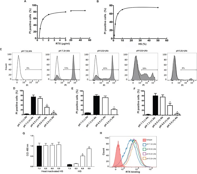
A. Raji cells (5×10⁵/100 μl) were treated with different concentrations of RTX (0.5 to 50 μg/ml) and incubated for 30 min at 37°C and pH 7.3, in RPMI medium supplemented with 10% of HS. Necrosis was then evaluated by propidium iodide staining and flow cytometry. A representative experiment (n=6) is shown. B. Raji cells (5 × 10⁵/100 μl) were treated with 2 μg/ml of RTX and incubated in RPMI medium supplemented with different concentrations of HS (1 to 50%) for 30 min at 37°C and pH 7.3. Then, necrosis was evaluated. A representative experiment (n=8) is shown. C and D. Raji cells (5 × 10⁵/100 μl) were treated with RTX (2 μg/ml) and then were incubated in RPMI medium supplemented with untreated (grey histograms) or heat-inactivated HS (open histograms) (10%) for 30 min at 37°C, at different values of pH. Then, necrosis was evaluated by propidium iodide staining and flow cytometry. Representative histograms and the mean ± SEM of 5 experiments performed in duplicate are shown (* p<0.05 vs pH 7.3 +Ab). E. Daudi cells (5 × 10⁵/100 μl) were treated with RTX (2 μg/ml) and then were incubated in RPMI medium supplemented with HS (20%) for 30 min at 37°C at different values of pH. Then, necrosis was evaluated by propidium iodide staining and flow cytometry. Data represent the mean ± SEM of 4 experiments performed in duplicate (* p<0.05 vs pH 7.3 +Ab). F. Raji cells (5 × 10⁵/100 μl) were treated with RTX (2 μg/ml) and then were incubated in Ca2+/Mg2+-veronal-buffered saline supplemented with 10% HS for 30 min at 37°C, at different values of pH. Then, necrosis was evaluated by propidium iodide staining and flow cytometry. Data represent the mean ± SEM of 4 experiments (* p<0.05 vs pH 7.3 +Ab). G. Raji cells (5 × 10⁵/100 μl) were treated with RTX (2 μg/ml) and then were incubated in RPMI medium supplemented with heat-inactivated or untreated HS (10%) for 30 min at 37°C, at different pH values. Then, cells were suspended in colourless RPMI and cell viability was analyzed using a colorimetric commercial kit based on the ability of viable cells to reduce a tetrazolium compound to a formazan product, which was quantified by absorbance at 490 nm. Data represent the mean ± SEM of 4 experiments performed in duplicate (* p<0.05 vs pH 7.3). H. Raji cells (5 × 10⁵/100 μl) were incubated at room temperature for 30 min with RTX (2 μg/ml) in culture medium supplemented with 10% of heat-inactivated HS adjusted to different pH values. Then, cells were washed, and the binding of RTX to Raji cells was revealed using a FITC-labeled mAb directed to the Fc fragment of human IgG and flow cytometry. A representative experiment is shown (n=4).