


FIG. 5.
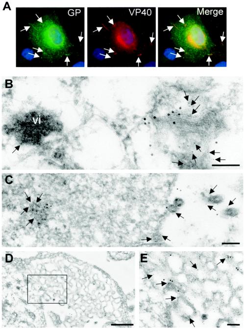
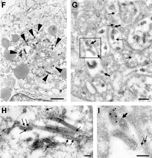
Distribution of GP and VP40 in MARV-infected cells. (A) MARV-infected Vero E6 cells were fixed at 2 days postinfection and immunostained with a rabbit anti-GP antibody and a mouse anti-VP40 antibody. Bound antibodies were detected using secondary donkey anti-rabbit IgG conjugated with FITC and donkey anti-mouse antibody conjugated with rhodamine. The nuclei were counterstained with DAPI (4′,6′-diamidino-2-phenylindole). Arrows show peripherally located clusters where GP and VP40 are colocalized. (B to E and H) MARV-infected Vero cells were fixed at 48 h postinfection, dehydrated, and embedded in LR Gold. Double immunostaining of ultrathin sections was performed with a rabbit anti-GP antibody (12-nm-diameter gold beads) and a mouse anti-VP40 antibody (6-nm-diameter gold beads) (arrows). vi, viral inclusion. (E) Enlarged framed fragment of panel D. (F, G, and I) MARV-infected human macrophages were fixed at 48 h postinfection, dehydrated, and embedded either in Epon (F) or in LR Gold (G and I). (F) MARV budding inside MVBs (arrowheads indicate viral particles). (G and I) Double immunostaining of ultrathin sections was performed with a rabbit anti-VP40 antibody (12-nm-diameter gold beads) and a mouse anti-Lamp-1 antibody (6-nm-diameter gold beads) (arrows). (I) Enlarged framed fragment of panel G. Thick arrows show Lamp-1-bound gold beads in the viral particles. Bars, 100 nm (B, C, E, H, and I), 500 nm (D), 1,000 nm (F), and 200 nm (G).