


Figure 5.
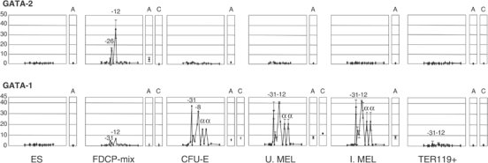
GATA-1 and GATA-2 bind to specific elements in the α-globin locus according to their expression pattern. Plots follow Figure 4. Above: GATA-2 binds only in FDCP-mix. Below: GATA-1 replaces GATA-2 as differentiation proceeds and binds most HSs, including the α-globin promoters. In late erythroblasts, no GATA binding is detected. A, HS2 of the β LCR; C, −2.8 region of the GATA-2 locus (Grass et al, 2003). Error bars represent ±1 s.d. from two independent ChIPs.