


Figure 6.
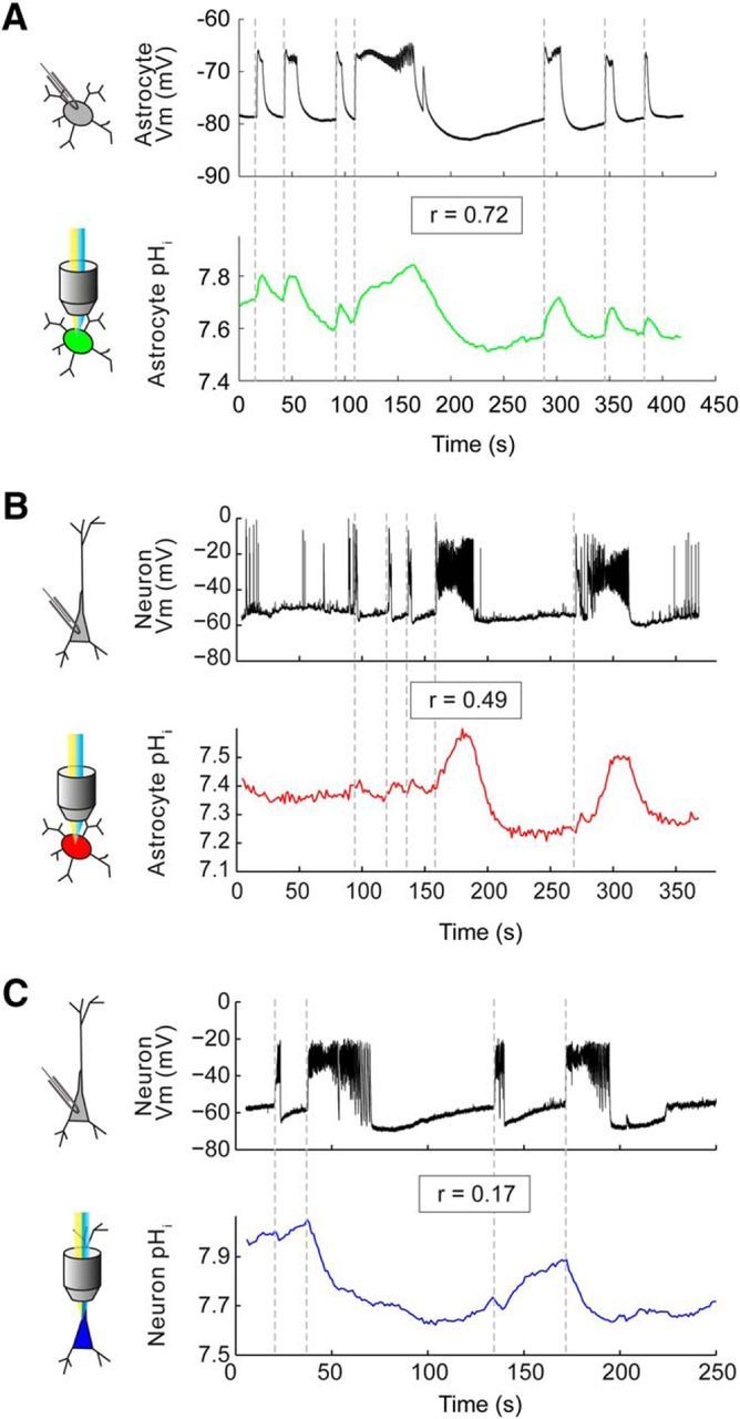
Simultaneous recordings from different cell-pair combinations reveal that astrocytic pH dynamics closely reflect both astrocytic and neuronal membrane potential dynamics. A, Paired pH imaging (green trace) and whole-cell patch-clamp recording (black trace) from a pair of astrocytes in stratum radiatum of CA3 revealed a close correlation between changes in astrocytic pH and changes in astrocytic membrane potential during epileptiform activity (r = 0.72, Pearson correlation). B, A similar experiment on an astrocyte–neuron pair also revealed a high degree of correlation between changes in astrocytic pH and neuronal membrane potential (r = 0.49, Pearson correlation). C, In contrast, a similar experiment on a neuron–neuron pair revealed a much lower correlation between the change in neuronal pH and the change in neuronal membrane potential (r = 0.17, Pearson correlation).