


Figure 1. LAST1 was lowly expressed in GC tissues and cell lines.
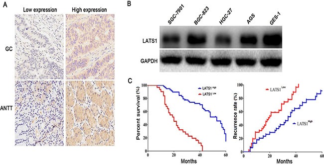
(A) Differential protein expression levels (low or high) of LATS1 were examined in GC tissues and ANTT by IHC staining (magnification, 200×). (B) Western blotting analysis of LATS1 expression in GC cell lines. (C) Low expression of LATS1 was markedly correlated with the overall survival and recurrence free survival of the GC patients. These data were analyzed using Kaplan-Meier survival analysis between the GC patients with low LATS1 expression and high LATS1 expression according to the immunostaining scores.