


Figure 4.
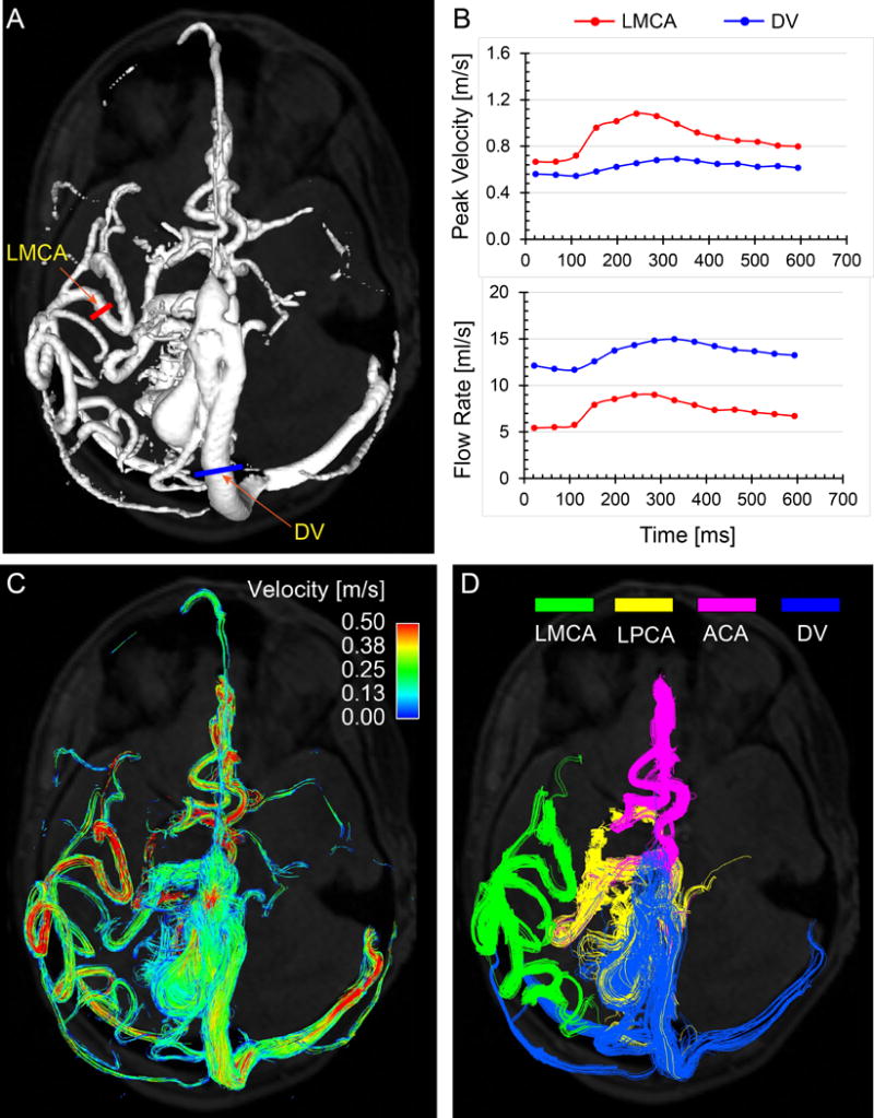
Illustration of 4D flow MRI for hemodynamic assessment in a patient with cerebral AVM (age 40 years, female, Spetzler-Martin grade = 4). A: 3D phase-contrast MR angiogram (PC-MRA) used for the orientation of 2D analysis planes. B: regional flow characteristics (peak velocity and flow rate) at user-defined vessel locations (examples for the feeding LMCA and the largest draining vein). C: Cumulative flow pathways of the entire cerebral vasculature over one cardiac cycle are depicted using time-integrated 3D pathlines which are color-coded according to local vascular velocity magnitude. D: selective vascular cartography illustrating connectivity and flow contribution of the major feeding arteries and draining vein. LMCA: left middle cerebral artery (green), LPCA: left posterior cerebral artery (yellow), ACA: anterior cerebral artery (pink), DV: draining vein (blue).