


Figure 5. Blocking of sirt1 activity inhibited the melatonin-induced protection against hydrogen peroxide in keratinocyte.
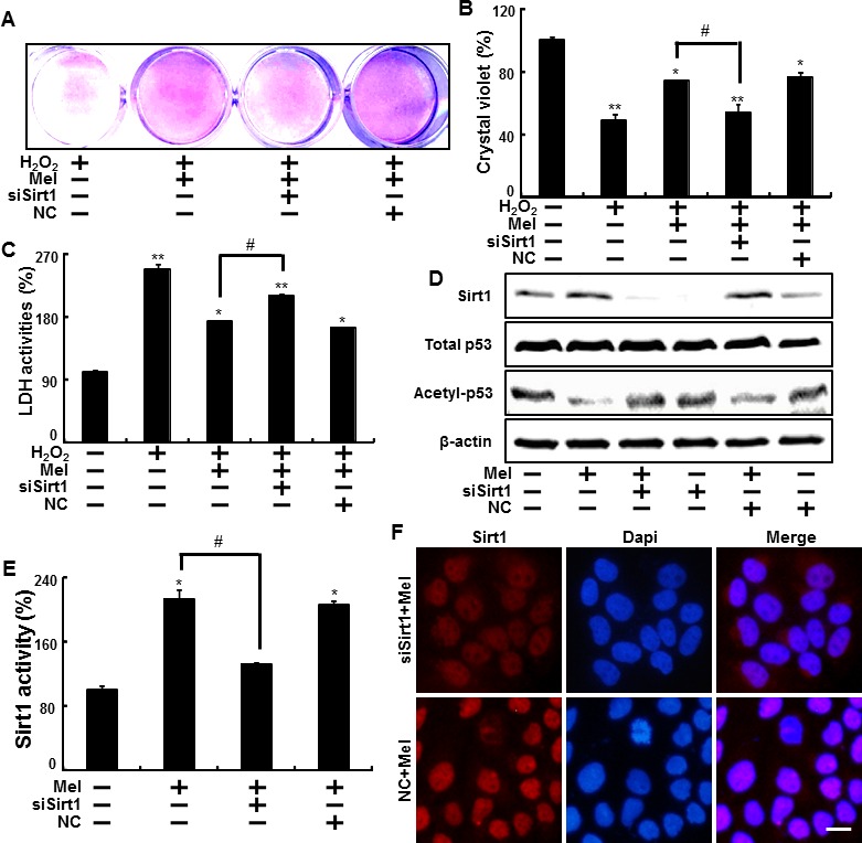
Sirt1 small interfering RNA (siSirt1) or negative control siRNA (NC siRNA) transfected HaCaT keratinocytes were incubated with 800 μM H2O2 for 18 hr in the presence of melatonin. The cell viability was measured by crystal violet staining. Viability of the control cells was set at 100%, and viability relative to the control was measured A., B. LDH activities were measured by quantifying LDH released in the medium C. siSirt1 or NC siRNA (negative control siRNA) transfected HaCaT keratinocytes were incubated with melatonin for 24 hr. Western blot for sirt1 and acetyl-p53 proteins was analyzed from HaCaT keratinocytes. β-actin was used as loading control D. Sirt1 deacetylase activities in nuclear were analyzed from HaCaT keratinocytes E. The cells were immunostained with sirt1 antibody (red) and observed in the fluorescent view Scale bar: 10 μm F. Bar graph indicates the mean ± standard error of the mean (SEM) (n = 4). * p < 0.05, ** p < 0.01 significant differences between control and each treatment group and # p < 0.01; significantly different when compared with H2O2 and melatonin-treated group.