


Figure 4.
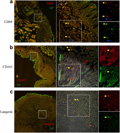
CD163+ and CD68+ monocytes/macrophages and Langerhans cells in the upper respiratory tract are virus antigen–positive. (a–c) These sections are representative samples of viral antigen (nucleoprotein (NP))-positive signals that are in the epithelium (a–c, red arrows), the cytoplasm or the intracellular compartment of CD68+ cells (a, yellow arrows and blue arrow), and the intracellular compartment of CD163+ cells (b, yellow arrows) and the cytoplasm of Langerin+ (c, yellow arrows). Pharyngeal mucosa tissue samples were stained for severe acute respiratory syndrome coronavirus (SARS-CoV) NP (fluorescein isothiocyanate) (a–c), CD68 (tetramethylrhodamine (TRITC)) (a), CD163 (TRITC) (b), and langerin (TRITC) (c). The left panel shows a low magnification overview (× 100). The middle panel shows a higher magnification of the boxed area in the left panel (× 400). The right panel shows the single color of the boxed area in the middle panel.