


Figure 3.
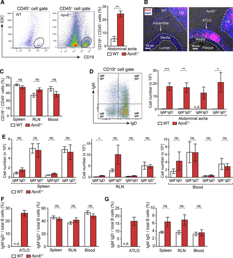
Artery tertiary lymphoid organs (ATLOs) harbor a diverse repertoire of B-cell subsets. A, Flow cytometry of CD19+ B cells of total CD45+ leukocytes in abdominal aorta of 80-week-old wild-type (WT) and ApoE−/− mice (WT, n=11; ApoE−/−, n=10). B, Immunofluorescence staining with anti-B220 antisera shows B cells in ATLOs and lymph nodes (LNs) but none in the media (M) or plaque (P) or in WT adventitia. C, B cells in the spleen, renal LNs (RLNs), and blood of WT and ApoE−/− mice (WT, n=8; ApoE−/−, n=6). Flow cytometric analysis of IgM+IgD−, IgM+IgD+, IgM−IgD−, and IgM−IgD+ B cells per total CD19+ B cells in abdominal aorta (D) and spleen, RLN, and blood of 80-week-old WT and ApoE−/− mice (WT n=4; ApoE−/− n=5; E). Percentages of IgM+IgD+ B cells (F) and IgM−IgD− B cells per total B cells in ATLOs, spleen, RLN, and blood of age-matched WT and ApoE−/− mice (G). Results represent mean±SEM; *P<0.05, **P<0.01, and ***P<0.001; 2-sided unpaired Student t test. n indicates the number of experiments; n.d., not detectable; ns, not significant; and SSC, side scatter.