


Figure 2. Architecture of the HKU1 S1 subunit.
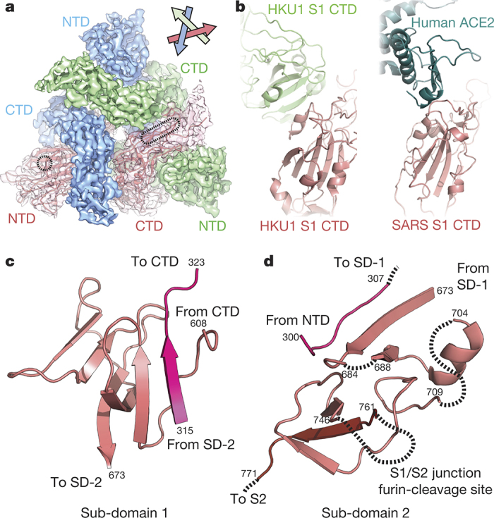
a, EM density corresponding to each S1 protomer is shown. The putative glycan-binding and protein-receptor-binding sites are indicated with dashed shapes on the NTD and CTD, respectively. b, The HKU1 S1 CTD forms quaternary interactions with an adjacent CTD using a surface similar to that used by SARS CTD to bind its receptor, ACE2 (ref. 11). c, Sub-domain 1 is composed of amino acid residues before and after the S1 CTD. d, Sub-domain 2 is composed of S1 sequence C-terminal to the CTD, a short peptide following the NTD, and the N-terminal strand of S2, which follows the S1/S2 furin-cleavage site.