


Figure 2.
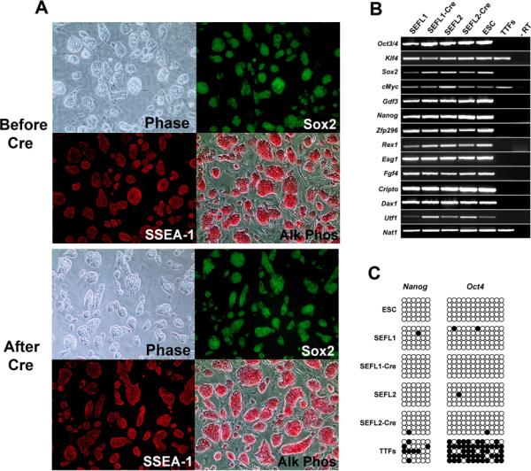
Characterization of iPS clones before and after STEMCCA-loxP excision. (A) Representative images of iPS cells derived using the constitutive STEMCCA-loxP vector before and after Cre-mediated excision show normal colony morphology (Phase), Sox2-GFP reporter gene expression, SSEA1 positive immunostaining, and robust alkaline phosphatase activity (Alk Phos). (B) Expression of ESC `marker' genes detected by RT-PCR in iPS cell clones before (SEFL1 and SEFL2) and after excision (SEFL1-Cre and SEFL2-Cre). Representative samples from murine ESC and unmanipulated tail tip fibroblasts (TTFs) are also shown for comparison. An iPS cell sample prepared without RT was used as negative control (-RT). (C) Promoter regions of Nanog and Oct4 genes were analyzed for methylation status using bisulfite sequencing. Similar to ESC, all iPS cell clones (SEFL1, SEFL1-Cre, SEFL2 and SEFL2-Cre) showed mostly unmethylated CpG motifs (open circles) in sharp contrast to parental TTFs, in which the extracted DNA was mostly methylated (closed circles).